

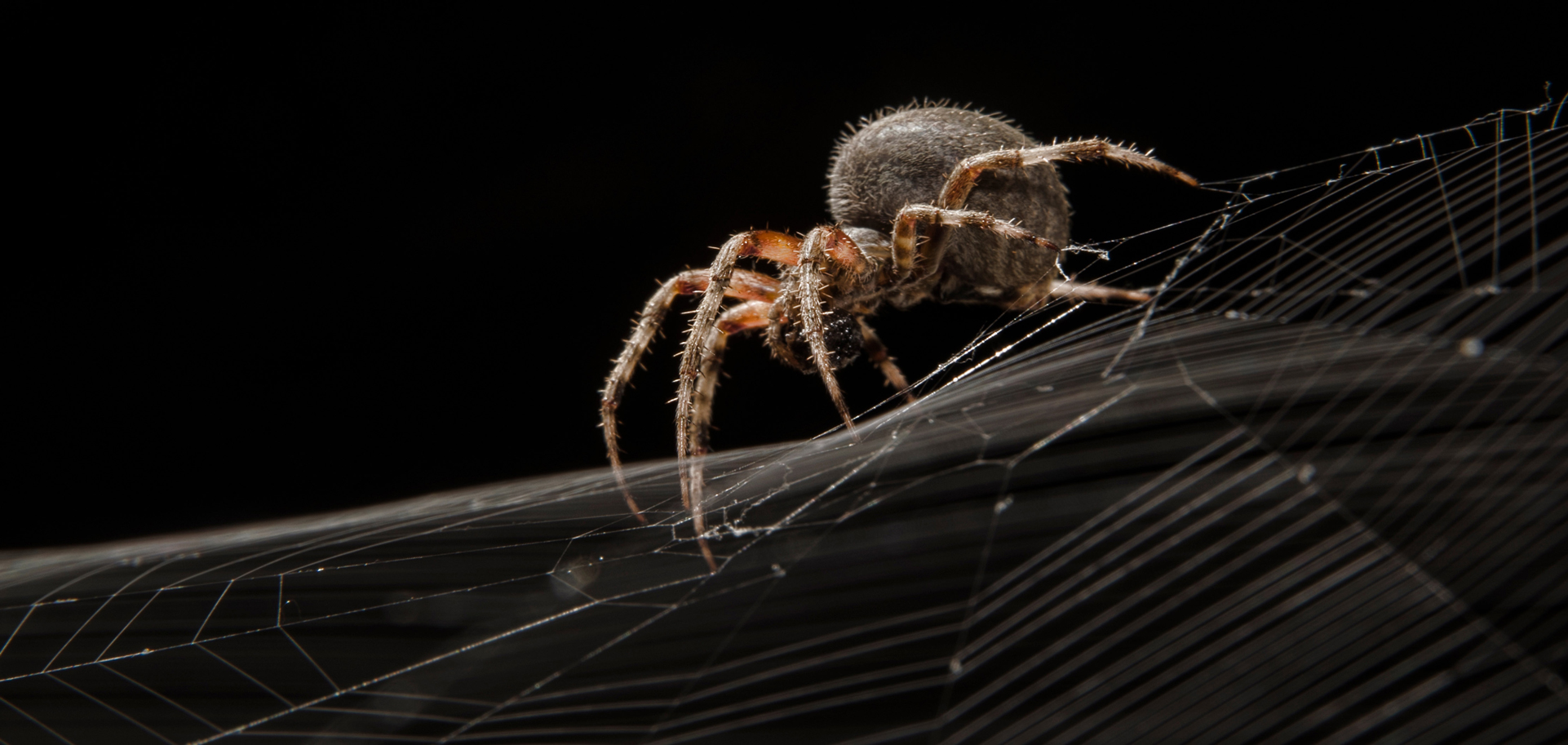
Spiders may induce spine-tingling chills, but they also serve as inspiration for medical advances. A unique, new glove fitted with spinneret-like devices releases ultrathin polymer fibers that can be spun directly onto wounds in real time. This device could be especially useful in battlefield environments, hospitals and sports arenas.
Ethereal ghosts, bodiless brains, spooky spiders, hairy wolves, slimy eyeballs, creepy cyclops, shadowy bats and severed limbs. What more could you want for Halloween? I’m guessing not much, and there’s a one-stop shop for all of it: science.
Almost every day, we get new evidence that science has one foot in the realm of the supernatural. For example, recently an elusive and declining animal—a “ghost,” you might say—has been found to still live in the Mediterranean Sea. A rare encounter with a juvenile great white shark (Carcharodon carcharias) has reignited scientific interest in this mysterious population of these apex predators, who still may be reproducing in the region.
And from mini brains to spider-inspired gloves to wolf-apple coatings, scientists are turning eerie-sounding experiments into real innovations that could revolutionize our health and sustainability efforts. Lab-grown brain organoids may replace animal testing, spider-silk gloves could create instant wound dressings and wolf-apple starch can keep veggies fresh. Even dreaded microplastics have been found lurking in human retinas—offering both wonder and a warning about the modern world.
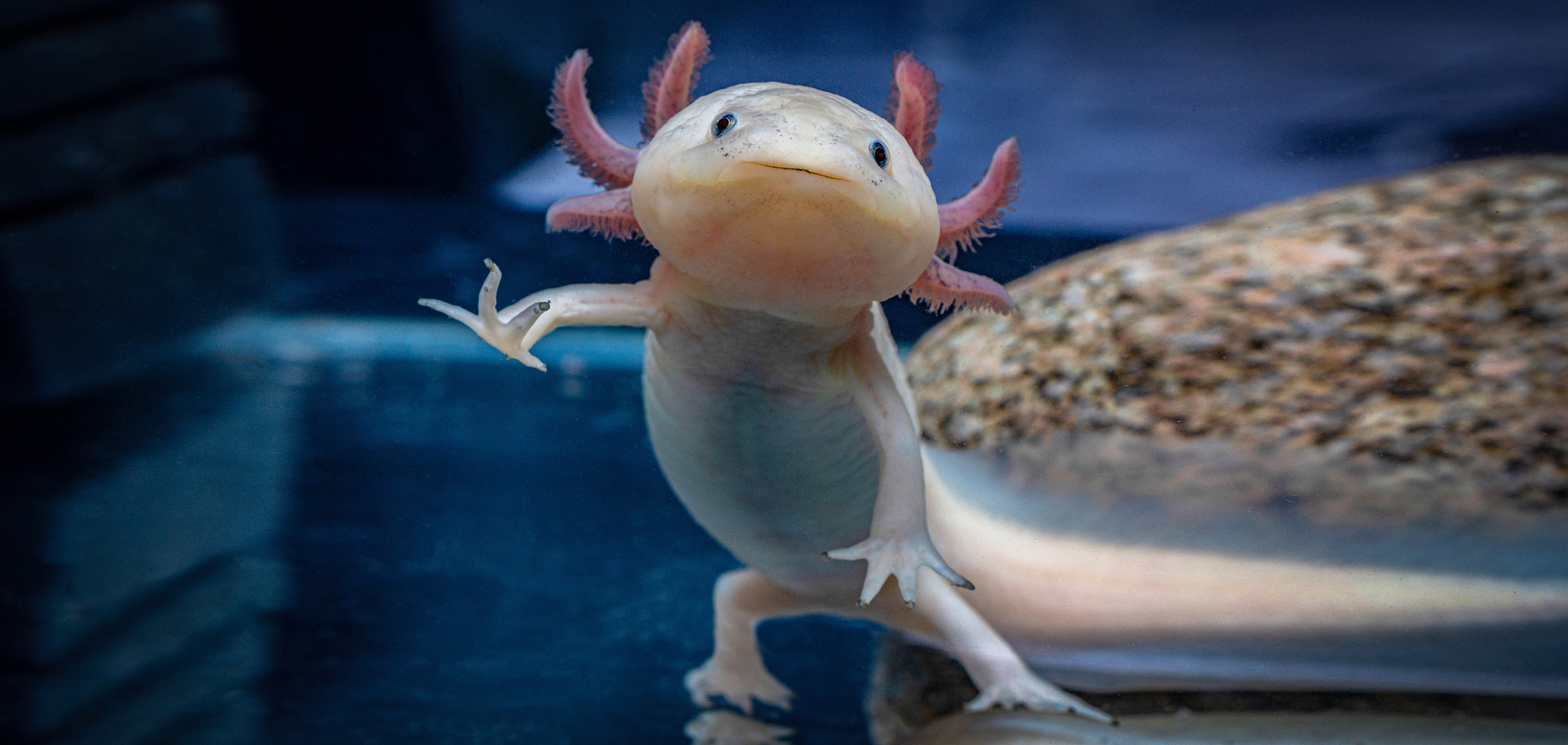
With their fascinating ability to regrow entire limbs and internal organs, axolotls are the ideal models for studying regeneration. Scientists have now found a factor that tells cells which part of an arm to regenerate—and used it to reprogram the identity of cells as they develop. This breakthrough has implications for tissue engineering.
A bizarre, cyclops-like creature from nearly 600 million years ago may hold the key to how your eyes—and even your sleep cycle—evolved. All vertebrates, including humans, trace their vision back to a single, light-sensitive “median eye” perched atop a worm-like ancestor’s head. As this ancient animal shifted from a sedentary to a more active lifestyle, it lost and then reinvented its vision, eventually giving rise to the paired, image-forming eyes we rely on today.
Bats, one of the most iconic symbols of Halloween, have inspired researchers to design flexible wings that boost lift and improve flight performance. Their invention could lead to more efficient drones or energy-harvesting technologies. And with their fascinating ability to regrow entire limbs and internal organs, Mexican axolotls are the ideal models for studying regeneration. Scientists have now found a factor that tells cells which part of the arm to regenerate—and used it to reprogram the identity of cells as they develop. This breakthrough for the regeneration research field has implications for engineering tissues, including those of humans.
A “ghost” reignites a Mediterranean mystery
A ghost lives in Mediterranean waters. Despite the widely held consensus that great white sharks have almost entirely disappeared off Spain’s coastline, scientists now say that a mysterious (or “ghost” population) has never fully vanished.
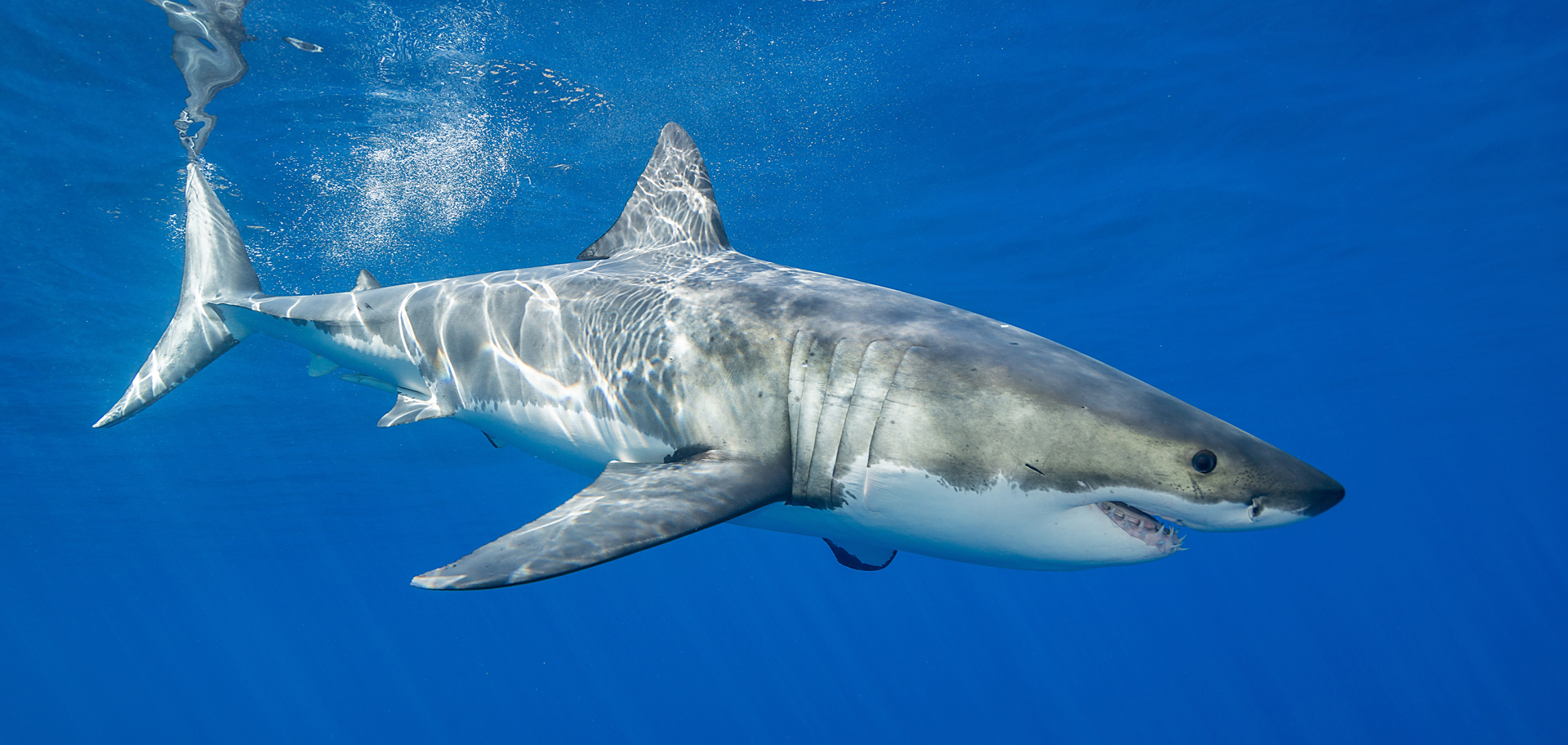
Scientists believe only a few hundred great white sharks remain in the Mediterranean, making them critically rare. Overfishing and illegal trade in North African markets have pushed the regional population toward the brink of extinction. At least 40 great white sharks were reportedly killed in 2025 along North African coasts.
One documentation occurred on April 20, 2023, when local fishermen accidentally caught a juvenile great white shark off the coast of the eastern peninsula. The young shark measured about 6.8 feet in length and weighed approximately 200 pounds. Encounters like this are extremely uncommon in the region, prompting scientists to take a closer look at historical records of reports dating back to 1862. They found that sightings are infrequent, but they show a consistent pattern over time. Rather than disappearing completely, the species quietly persists in the region. Finding younger sharks could indicate that breeding is still taking place somewhere in the Mediterranean, an idea that researchers are eager to investigate further.
Many people today fear great white sharks. To address the powerful emotions that the animals often inspire, the researchers—in their study that was published in the open access journal Acta Ichthyologica et Piscatoria in January 2026—referenced H. P. Lovecraft’s famous observation that “the oldest and strongest emotion of mankind is fear, and the oldest and strongest kind of fear is fear of the unknown.” Better scientific understanding can help change public perception, say the scientists. By shedding light on the biology and ecology of the great white shark, research can help replace unfounded myths with genuine understanding. Great white sharks have a fundamental role in marine ecosystems. As a highly migratory, pelagic species, they redistribute energy and nutrients across vast distances. They serve as nature’s scavengers; by consuming carrion, they keep ecosystems clean. Even in death, their descent to the seafloor provides a critical pulse of nourishment for deep-sea communities.
At present, the great white shark is classified as vulnerable on the IUCN Red List, and global numbers are believed to be declining. Long-term monitoring programs that combine direct sightings with modern tracking technologies are needed to better understand great white sharks in the Mediterranean and to help develop evidence-based conservation strategies to protect this iconic predator.
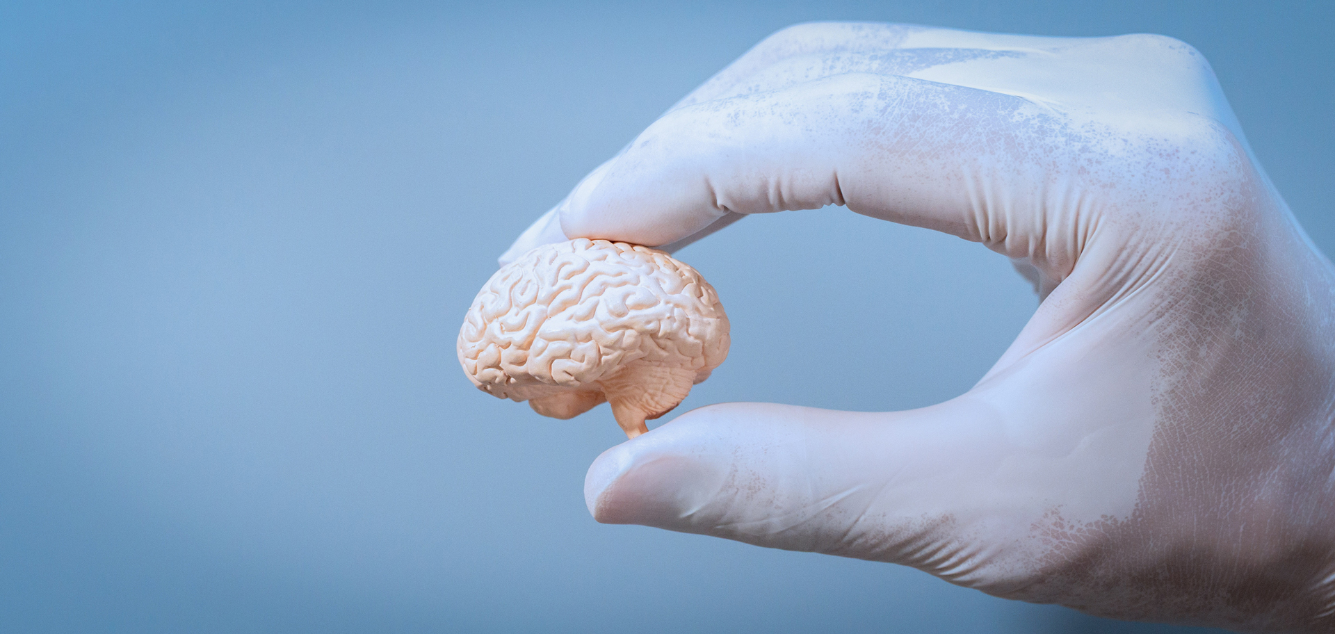
In August 2025, scientists successfully cultivated a three-dimensional, mini brain in a dish. Such an organoid could become a valuable tool for studying brain function—or, at least, serve as a “lab-grown lunch for a zombie.”
Creepy science saves lives
Things that sound like props from a horror movie are now being used in clever ways to improve human health. In one study that was described in the journal ACS Sensors in August 2025, scientists successfully cultivated a three-dimensional, mini brain in a dish. Over the course of two years, cultured human nerve cells multiplied and organized themselves into a functioning organoid capable of generating electrical activity. This advancement allows researchers to explore how brain cells interact and communicate without using animals in experiments. Future advances could make these organoids valuable tools for studying brain function; or, as the researchers jokingly note, a possible “lab-grown lunch option for zombies.”
Spiders were the inspiration for a unique, new glove fitted with spinneret-like devices that release ultrathin polymer fibers. The invention, described in the journal ACS Applied Materials and Interfaces in August 2025, allows medical workers to spin wound dressings directly onto injuries in real time. Such a system could be especially useful in battlefield environments, hospitals or sports arenas. And in case anyone is wondering, state the inventors, these experiments did not involve any radioactive spider bites.
In an article that appeared in the journal ACS Food Science and Technology in August 2025, scientists report that they’ve found that starch extracted from the wolf apple—a fruit native to Brazil and a favorite of the maned wolf—can be transformed into a natural, edible coating that helps preserve food. When applied to baby carrots, the coating kept them bright and fresh for up to 15 days at room temperature. The material offers a safe, cost-effective way to extend the shelf life of produce—even whether or not there’s a full moon.
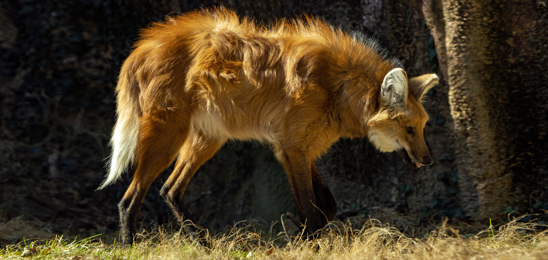
Residents of the open and semi-open grasslands, savannas and scrub forests of central and eastern South America, maned wolves are omnivores that get about 50% of their daily nutrition from plant matter, especially a tomato-like fruit called the “wolf apple.” The fruit makes up 40% to 90% of the wolves’ plant intake year-round.
After examining 12 postmortem human retinas, scientists discovered microplastic particles in every sample. The plastics varied in type and concentration, revealing how pervasive they have become, even in such delicate tissues. The researchers, who published their report in the journal ACS Environmental Science and Technology Letters in October 2025 say these findings lay important groundwork for future investigations into how microplastics might influence vision and overall eye health.
A shocking “cyclops” originated the human eye
Humans share a surprisingly strange ancestor with all other vertebrates. Far back in evolutionary history, one of our earliest relatives had a single eye—much like a cyclops—positioned on top of its head. All vertebrates can be traced back to this ancient, one-eyed organism; and the remains of that original “median eye” still exist today, but in a very different form.
Researchers from England’s University of Sussex and Sweden’s Lund University, who published their findings in the journal Current Biology in February 2026, say this distant ancestor lived nearly 600 million years ago. It was a small, wormlike animal that spent most of its time in one place, feeding by filtering plankton from seawater. Earlier in its evolutionary history, it likely had two eyes, similar to many other animals. While it’s not known whether the paired eyes in our branch of the evolutionary tree were just light-sensitive cells or simple image-forming eyes, we do know that the organism later lost them. As the creature adapted to a more stationary lifestyle, having two eyes was no longer necessary. Over time, those paired eyes disappeared.
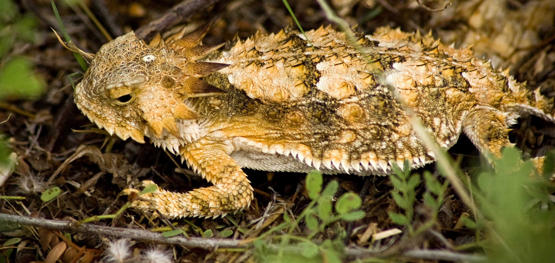
Regal horned lizards are found natively in the arid and semiarid deserts of southern Arizona, southwestern New Mexico and western Mexico (including Sinaloa and Sonora). The light spot in the middle of the back of the head of the animal forms the median eye and creates the semblance of a cyclops. ©brdavids, Wikimedia Commons
Even after losing its paired eyes, our ancestor retained a cluster of light-sensitive cells in the center of its head. These cells gradually formed a simple, single eye that could detect light and darkness and help the animal sense orientation. Millions of years later, the organism became more active again and returned to a swimming lifestyle. That shift created new pressure to develop better vision. Researchers believe that parts of the original median eye eventually gave rise to new, paired eyes capable of forming images.
This unusual evolutionary path helps explain why vertebrate eyes are built differently from those of other animals like insects and squid. The film of our eyes—the retina—developed from the brain, whereas the eyes of insects and squid originated in the skin on the sides of the head. In vertebrates, the retina is essentially an extension of the brain. In contrast, many other animals develop eyes from surface tissues on the body.
Researchers describe this history as an unusual evolutionary detour. Instead of evolving directly from earlier eye structures, vertebrate vision appears to have been rebuilt from this single median eye after the loss of earlier paired eyes. This conclusion comes from detailed comparisons of light-sensitive cells across different animal groups, as well as their structure and location within the body.
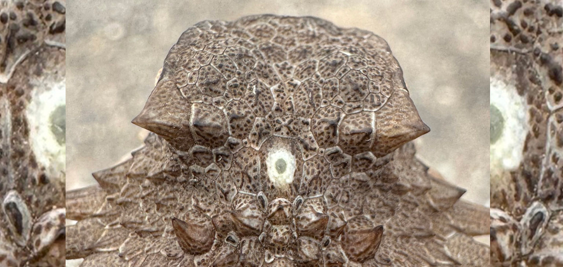
Often appearing as a pale, translucent scale on the top of the head, the parietal (or pineal) eye is a small, rudimentary photoreceptive organ. It cannot form clear images like regular eyes, but it senses changes in light, helping the lizard regulate its body temperature and track the time of day. ©Bruno Frias Morales, Wikimedia Commons
One of the most intriguing discoveries is that a piece of this ancient eye still exists in humans today. It has evolved into the pineal gland, a small, light-sensitive organ deep in the brain. The pineal gland produces melatonin, a hormone that helps control the body’s circadian rhythm, including sleep patterns. It’s mind-boggling, write the researchers, that our pineal gland’s ability to regulate our sleep according to light stems from the cyclopean median eye of a distant ancestor 600 million years ago.
Deformable bat wings portend the future
Bats have been a symbol of Halloween since its origins. The ancient Celtic festival of Samhain, meaning “summer’s end,” is the precursor to Halloween. Autumn bonfires lit during Samhain attracted swarms of flying insects, which in turn drew hungry bats out into the night skies.
In 1934, French entomologist Antoine Magnan wrote that bumblebees “should not be able to fly,” as their small wings should theoretically not be able to produce enough lift. It took modern high-speed camera technology to uncover what allowed airborne insects to fly: the leading-edge vortex. This phenomenon occurs when airflow around the leading edge of flapping wings rolls up into a vortex, creating a low-pressure region that boosts lift.
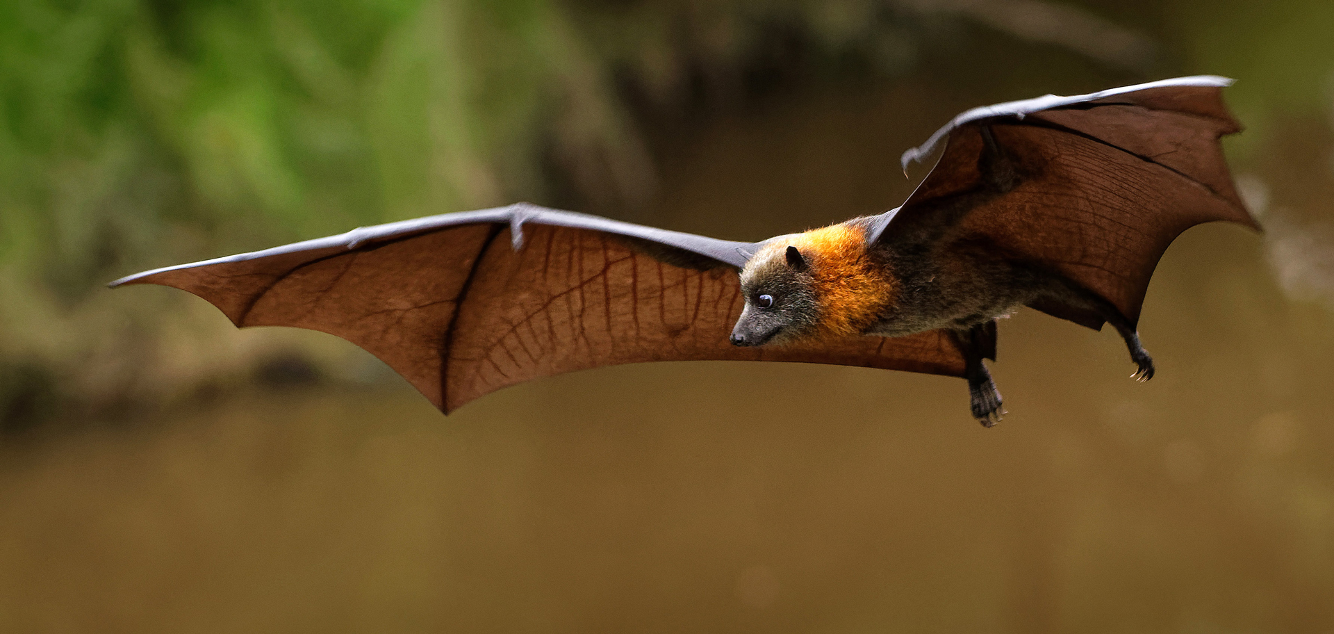
The gain in lift that flying bats achieve comes from the airflow that follows the smooth curvature of their membrane wings. Not only do wings have to be curved, but they have to be arced by just the right amount, as a wing that is too flexible doesn’t perform well.
On the other hand, bats—with their flexible membrane wings—are able to fly just as well as insects, if not more efficiently. In fact, some bats have been found to expend as much as 40% less energy than moths of a similar size. Researchers in the Unsteady Flow Diagnostics Laboratory at the Federal School of Technology in Lausanne, Switzerland, set out to study the aerodynamic potential of more flexible wings using an experimental platform with a highly deformable membrane made from a silicone-based polymer. They found that instead of creating a vortex, the air flows smoothly over the curved wings, generating more lift and making them even more efficient than rigid wings of the same size.
The researchers say that they know that bats hover and that they have deformable membrane wings. How the wing deformation affects the hovering performance is an important question, but doing experiments on live animals is difficult. So, they mounted a flexible membrane onto a rigid frame with edges that rotate around their axes. To help visualize the flow around the wing, they immersed their device in water mixed with polystyrene tracer particles. This allowed the scientists to indirectly alter the front and back angles of the wing, so they could observe how they aligned with the flow. Due to the membrane’s deformation, the flow wasn’t forced to roll up into a vortex; rather, it followed the wing’s curvature naturally without separating, creating more lift.
By using a simplified bio-inspired experiment, state the researchers in their paper published in the journal Proceedings of the National Academy of Sciences in January 2025, we can learn about nature’s fliers and how to build more efficient aerial vehicles. For example, as drones get smaller, they are more strongly affected by small, aerodynamic perturbations and unsteady gusts than larger vehicles like airplanes. Standard quadrotor drones might stop working with small disruptions, so one solution could be to use the same flapping wing motions as animals to build improved versions of these flyers that can hover and carry a payload more efficiently.
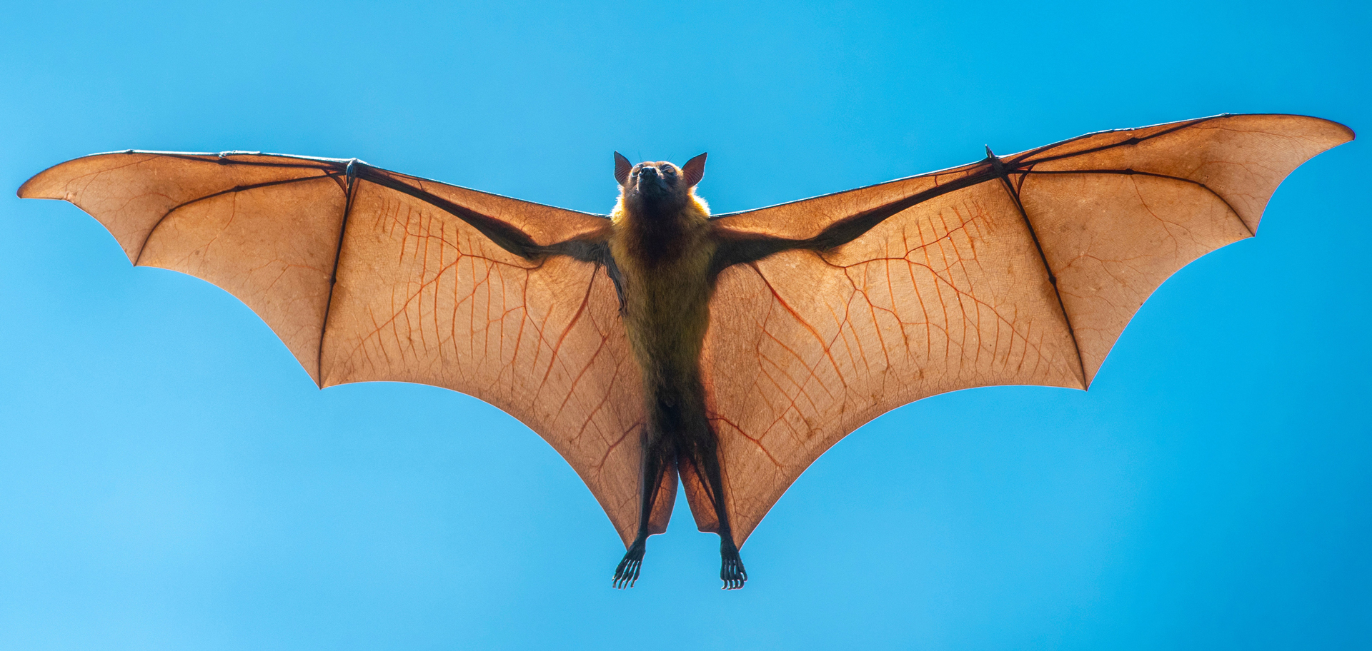
Nature’s fliers can teach us how to build more efficient aerial vehicles. For example, as drones get smaller, they are more strongly affected by minor aerodynamic perturbations and unsteady gusts of wind. Under such conditions, standard quadrotor drones can stop working. One solution is to mimic the same flapping wing motions that bats use.
These findings could also be used to upgrade existing energy technologies like wind turbines or to commercialize emerging systems like tidal harvesters that passively harness energy from the ocean’s currents. Advances in control technology and sensors, potentially combined with artificial intelligence, could enable the precise handling required to regulate the deformation of flexible membrane wings and adapt the performance of such flyers to varying flight missions or weather conditions.
Missing limbs regrow
Living in a murky lake around Mexico City, surrounded by aggressive and cannibalistic neighbors, the axolotl lives at constant risk of losing a limb to a neighbor’s nibble. Fortunately, lost limbs regrow and are functional in as few as eight weeks. To achieve this feat, the regrowing body parts must “know” their position within the axolotl body to regenerate the right structure for that specific location.
A long-sought code that tells the cells where they are located and gives body parts their identity has now been cracked by researchers at the Institute of Molecular Biotechnology of the Austrian Academy of Sciences. The study, published in the journal Nature in May 2025, shows how cells “remember” their position and, upon injury, switch on a signal that is broadcast across the one side of the limb and instructs cells to regenerate structures that match with their location.
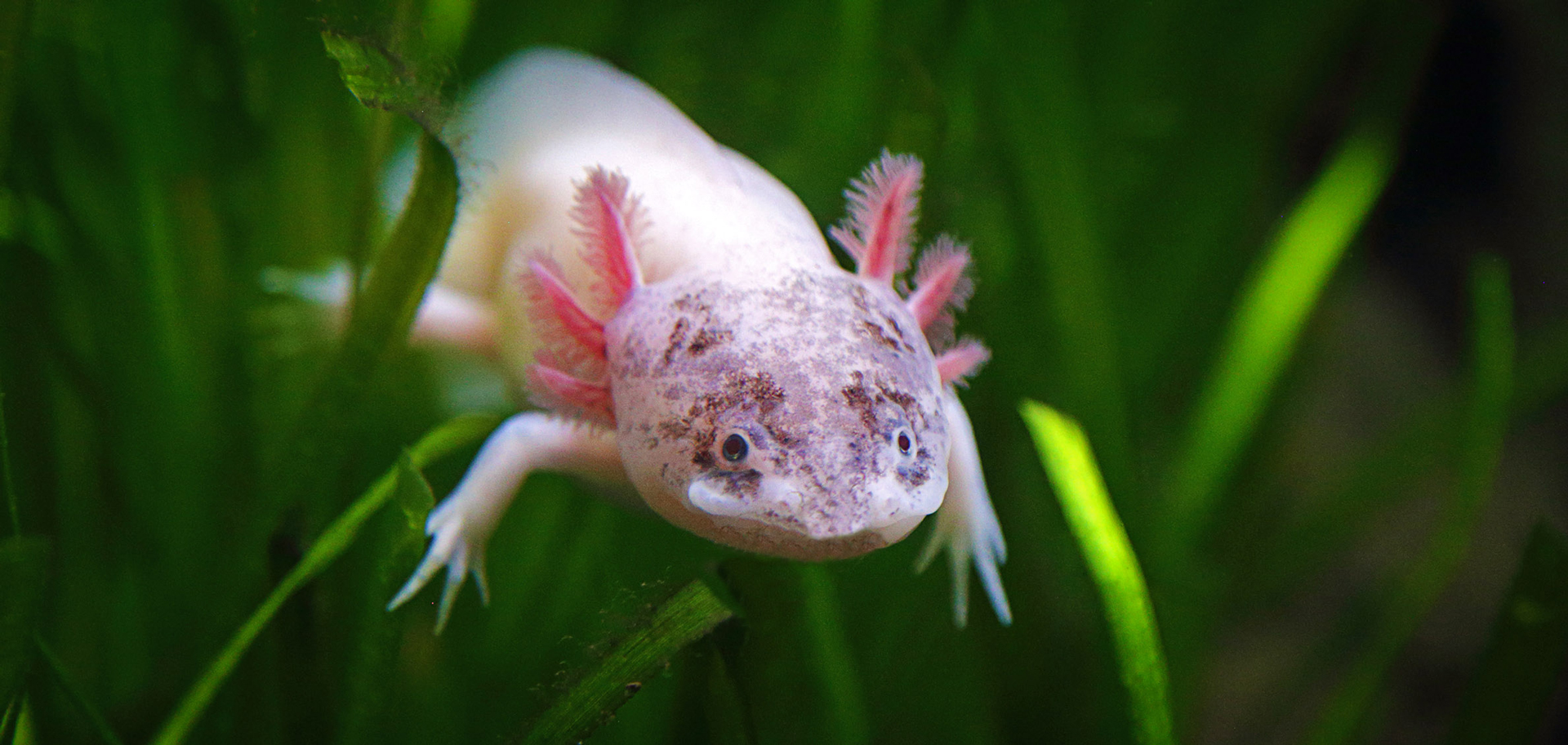
World Wildlife Fund calls the axolotl “the Peter Pan of salamanders.” While most amphibians grow out of their aquatic phase to begin their lives on land, the axolotl largely retains its larval characteristics and spends its adult life in the water.
Once regeneration starts, stem cells on the anterior (thumb) side express the signaling factor FGF8, while posterior (pinky) side stem cells express Shh. These two signals reinforce each other to tell cells to grow and pattern the regenerating arm. But axolotls have very large and complex genomes, and genetic tools readily available for other model organisms often do not exist for axolotls. Only recent advances with these molecular tools allowed the scientists to systematically search for signaling cues at the root of regeneration.
To their surprise, the researchers found hundreds of factors that differed in the anterior versus the posterior half of the limb even before an injury. But one gene stuck out: the appropriately named “Hand2” is only expressed on the posterior side and not at all in the anterior half. Experiments on developing and regenerating limbs confirmed an important role of Hand2 in switching on Shh after injury, demonstrating its role as a central player in providing positional information.
The scientists arrived at a new model for limb regeneration, resembling a radio broadcast: in the fully developed limb, only cells on the posterior side express Hand2 at low levels. This Hand2 expression maintains the cells’ stable memory of being in the pinky zone. Upon injury, these cells dial up Hand2 expression to a higher level, which switches on the Shh signal in a subset of Hand2-expressing cells. Shh cells near the Shh source then regenerate in the form cells with posterior-part identity; cells far away from the Shh signal regenerate as anterior-part cells. Once the limb is fully regenerated, cells again express Hand2 at a low level, ensuring a stable memory of their position is ready for the next cycle of injury and regeneration. These findings can, for the first time, explain how a preexisting positional memory signal reactivates upon injury to repeatedly induce correct patterning.
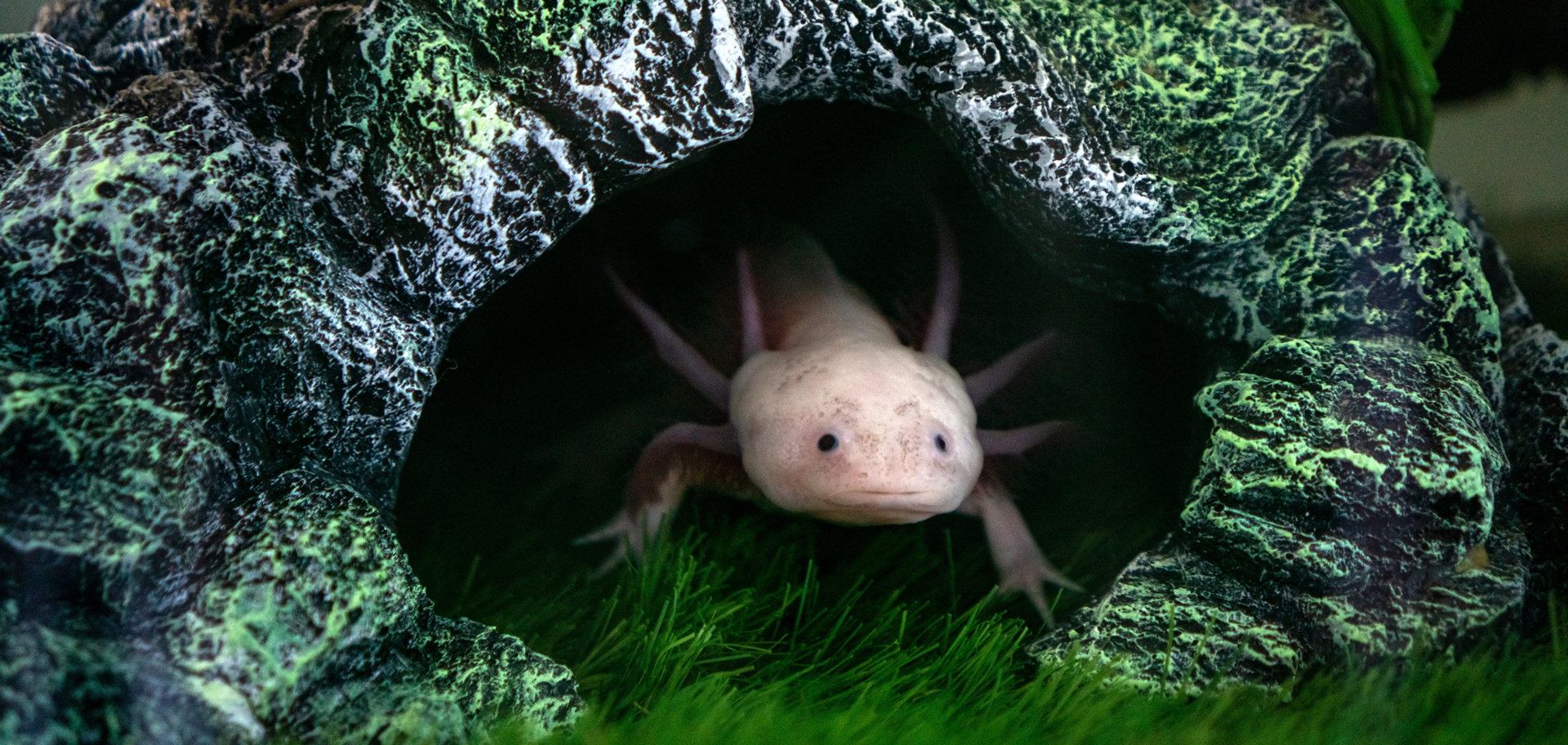
While commonly found in household aquariums and research labs, axolotls are nearly extinct in the wild, mainly due to habitat degradation, pollution and non-native predators in their limited geographic range. They are found in the wild in only one place on Earth: Lake Xochimilco (and the surrounding canal system) near Mexico City, Mexico.
The discovery, made possible by genetic manipulation and recently developed cell-tracing tools, is a major breakthrough for the entire regeneration field. The researchers say their model predicted that switching cells from an anterior identity to a posterior identity would be possible by taking advantage of the Shh broadcast. Indeed, when the researchers placed cells from the arm’s thumb side into the pinky side, these thumb cells regenerated and behaved like pinky cells as they fell into the range of the Shh broadcast.
The ability to alter cell identities holds immense potential for tissue engineering and regenerative therapies. This concept—essentially reprogramming cells to change their function—could enable scientists to transform cells in different parts of the body.
Knowing the signals that can transform cell identity and change their regenerative outputs and harnessing such signals might allow us to push cells beyond their normal biological limits. This could allow them to take on entirely new roles—an exciting prospect for medical innovation, conclude the researchers. The discovery that the axolotl relies on the Hand2-Shh signaling circuit for limb regeneration is particularly promising; these same genes are also present in humans. The fact that the axolotl reuses this circuit during adult life to regenerate a limb suggests that, if similar memory exists in human limbs, scientists may one day be able to target them to unlock new regenerative capabilities. Moreover, by expressing this gene in areas where it is not typically active, such as the anterior half of the limb, it could direct cells to initiate limb formation from scratch, fueling optimism that by using Hand2 expression along with other insights from the axolotl model, we may eventually be able to regrow limbs in mammals.

Science can be fascinating, strange and genuinely terrifying when we look into all of its corners. Once a year, it’s fun to examine the shocking, spooky and spine-chilling side of science.
Science steers advancements
Science can be fascinating, strange and genuinely terrifying when exploring biology, nature, physics and technology. In a very large part, though, scientific research fundamentally drives human progress by improving health, spurring innovations and expanding our understanding of the world.
Once a year, however, it is fun to look at the shocking, spooky and spine-chilling side of science.
Here’s to finding your true places and natural habitats,
Candy