

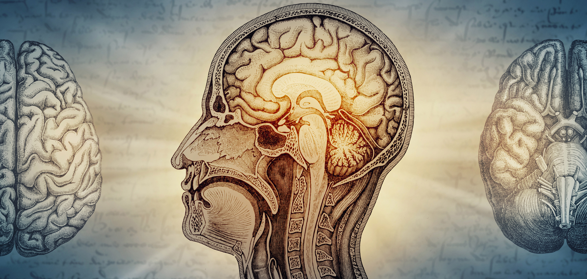
One long-standing cliche about human brains is that they evolved from an ancient “lizard brain,” with newer, rational layers getting stacked on top. But a tug-of-war in the brain’s wiring overturns that myth—and reveals a smarter way to build AI.
One of the scariest movies I have ever seen is the 1968 film Night of the Living Dead. In 1985, it was followed by the flick Return of the Living Dead. That movie created the pop-culture trope that zombies specifically crave and shout for brains. So, as we approach Halloween, I thought it would be fun to take a look at some brains—not only ours, but a few from other animals.
Speaking of tropes, one long-standing cliche about human brains is that they evolved from an ancient “lizard brain,” with newer, rational layers getting stacked on top. But we’re now learning that evolution, instead, appears to balance two competing styles of brain wiring, expanding one while shrinking the other depending on what an animal needs to survive.
Turning our attention to the insect world, leafcutter ants (Atta cephalotes) may not have what we call “lizard brains,” but they do have malleable ones. These insects live in highly organized colonies where every ant has a job. Recently, however, researchers found that they can flip those jobs like a switch. By manipulating just two neuropeptides, scientists can turn defenders into nurses or gardeners into leaf harvesters. These same molecular signals echo in naked mole-rats, revealing a deep evolutionary link in how complex societies function, even across species. The study also teases out a possible connection to insulin and longevity, hinting at new frontiers in understanding human behavior and lifespan.
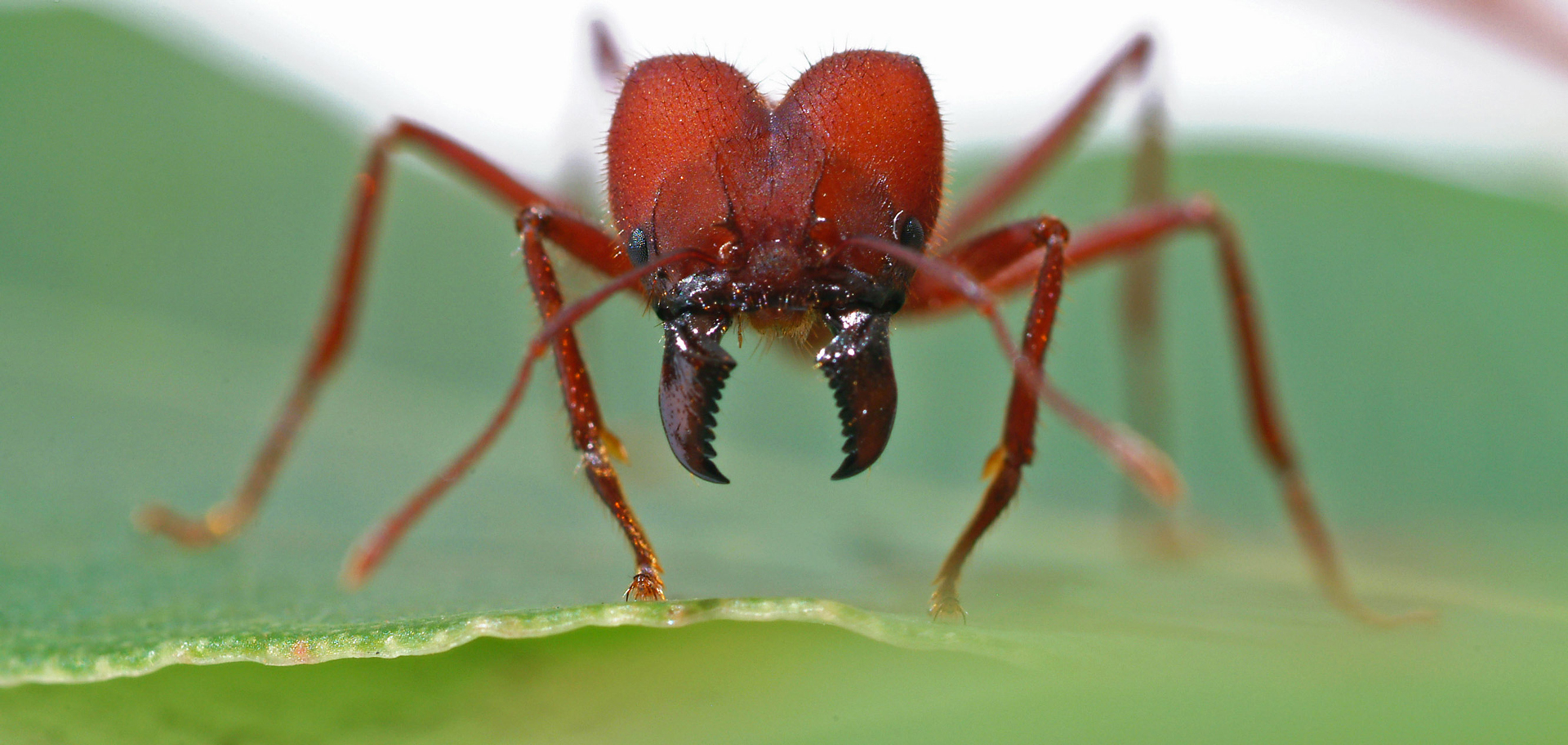
In eusocial superorganisms like leafcutter ant colonies, labor is divvied up according to body shape and size. Molecular signals can override that blueprint, however. Simple neuropeptides can reprogram ant behavior, reshuffling roles in nature’s most disciplined workforce.
Bees, too, have adaptable brains. Specific brain circuits help decide which jobs bees perform as they age. When those circuits are switched off, older worker bees begin caring for the queen again instead of carrying out their normal later-life duties. This means that a bee colony’s complex division of labor may be guided by neural switches hidden inside each bee’s brain.
And it’s not only zombies that may be coming for our brains. Tiny plastic particles may be quietly threatening our brain health. Microplastics—now widely found in food, water and even household dust—could trigger inflammation and damage in the brain through multiple biological pathways. Climate change, too, poses a threat: by adversely affecting the weather, it is likely to negatively influence the health of people who already have brain conditions.
Our brains are powerful come-back organs, however. They can be coaxed into suppressing pain by our simply feeling like we’re in nature. Brain scans confirm that the more present we feel in a 360-degree, virtual reality (VR) nature experience—like a forest or a waterfall scene—the stronger the pain-relieving effects.
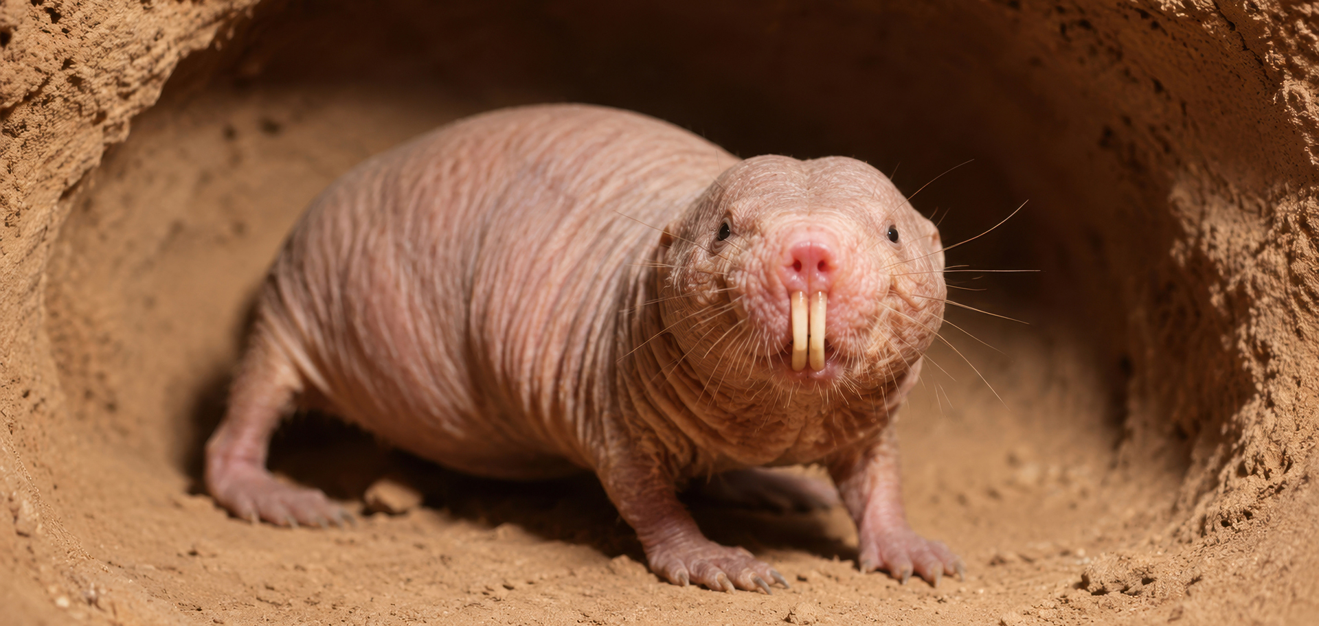
The same molecular signals in leafcutter ants echo in naked mole-rats, revealing a deep evolutionary link in how complex societies function, even across species.
Challenging the 70-year-old “lizard brain” myth
During our lives, many important choices seem to pit emotion against reason. Popular culture often describes that tension as a contest between two parts of the brain: an ancient “lizard brain” that runs on instinct and a newer, more advanced center for rational thought. But the evolutionary history of the brain appears to be far more complicated than a simple struggle between old and new.
In the 1950s, the theory that the brain evolved in layers starting with basic bodily functions, to emotions in the reptilian brain, leading up to sophisticated reasoning in humans was proposed. But research from scientists at Georgia Tech’s Institute for Neuroscience, Neurotechnology and Society (INNS) that was published in the journal Science Advances in April 2026 suggests that brain evolution may be better understood in terms of wiring rather than newer regions being stacked on top of older ones.
By examining the organization of both biological brains and artificial neural networks, scientists found evidence that evolution may involve allocating a limited amount of brain space among competing wiring strategies. It’s like there’s a computational tug-of-war between two fundamentally different kinds of neural organizations, both of which are established even before birth, state the researchers. These findings could help explain a long-running puzzle in brain evolution and may also point toward new ways of building AI systems that use less data and energy.
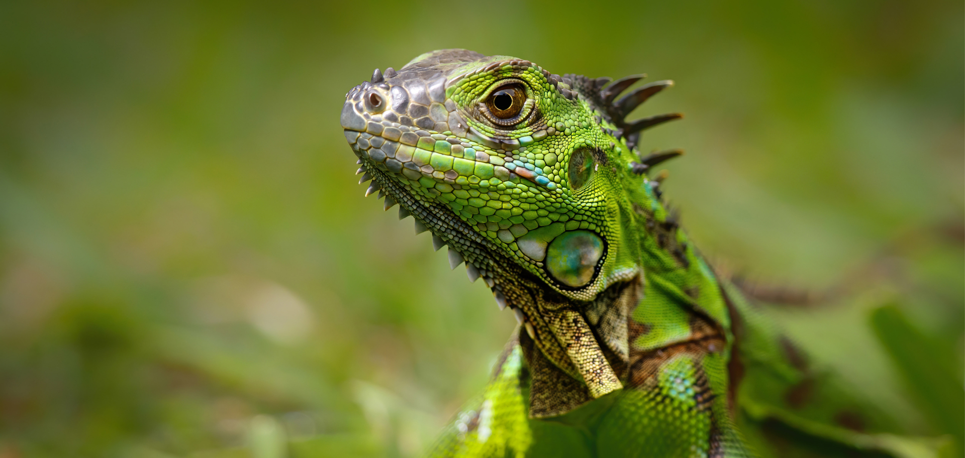
The “lizard brain” is a popular nickname for the oldest, most basic survival parts of the human brain, though neuroscientists consider it an outdated way to describe how real human brains work. Even iguanas don’t have “lizard brains”; their brains aren’t primitive—just uniquely adapted.
The reason why the lizard brain model falls short is that terms such as logical brain and lizard brain actually refer to groups of brain regions with very different functions. The neocortex (or logical brain), often associated with higher-level thought, forms the outer layer of the brain and is involved in perception, reasoning, vision and other complex abilities. The limbic system (or lizard brain) is harder to define so neatly.
Broadly speaking, the limbic system controls emotion; but it also has other components with distinct functions. The system contains separate regions involved in emotional regulation, memory, navigation and smell. The INNS researchers compared how this system changes across species. Rather than looking at individual regions one at a time, they examined how the limbic system and neocortex vary together over evolutionary history. When they did that, a clear pattern emerged. When one part of the limbic system was relatively large, the other limbic regions also tended to be larger. At the same time, the neocortex was generally smaller. That suggests the regions are not evolving independently. Rather, it’s a coordinated expansion of these regions across species. The pattern points to the limbic system behaving more like an integrated network than a collection of unrelated structures. Across evolution, its different components appear to expand and contract together.
The next question is what might cause that coordinated shift. The answer centers on the way different brain systems are wired before birth. In the neocortex, neural circuits are arranged as spatial maps. Brain regions that process nearby parts of the body, such as the index finger and thumb, are also located near one another. A similar spatial organization appears in systems that handle sight and sound.
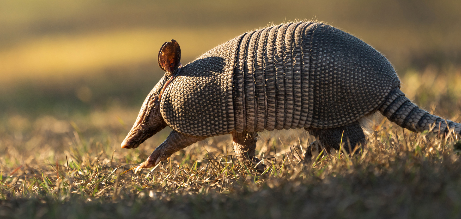
A nine-banded armadillo heavily depends on smell. The animal has a very large limbic system—a group of connected structures in the brain that manage emotions, memory and basic survival behaviors.
The limbic system, however, is organized differently. Instead of being laid out spatially, its wiring works more like a barcode, with distributed patterns of activity representing particular smells or complex memories. The researchers used artificial intelligence (AI) models to test whether these differences arise from built-in architecture or are mainly learned through experience. When an AI network was created with localized, spatial connections, it was naturally well suited for processing vision, sound and touch. In contrast, distributed barcode-style networks were necessary for strong performance on memory and smell recognition.
The researchers then investigated why the relative size of these brain systems changes so consistently among species. The basic idea is that the brain has limited resources. Energy and space are finite, so natural selection may favor whichever wiring system is most useful for survival in a particular environment. To test this, the team created a multimodal artificial network in which spatial (as in the neocortex) and distributed systems (as in the limbic system) competed for “real estate.” When the simulated environment rewarded smell, every region within the limbic system expanded while the neocortex became smaller. When vision was favored instead, the pattern reversed.
This trade-off may help explain striking differences between animals. The nine-banded armadillo, which depends heavily on smell, has a very large limbic system. The squirrel monkey, which relies strongly on vision, has a brain dominated by the neocortex. Across the 182 species included in the study, the findings suggest that brain evolution is less about adding progressively newer layers of “logic” and more about shifting space between different wiring systems according to what helps an animal survive.
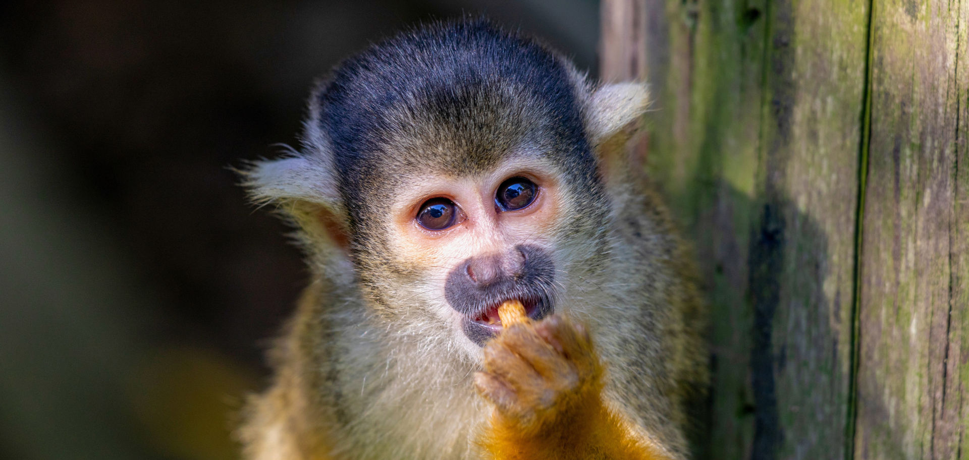
Squirrel monkeys possess one of the largest brain-to-body-mass ratios of any primate species. They rely strongly on vision; their brains are dominated by the neocortex.
The same principle may have implications beyond biology. If engineers can reproduce some of this built-in neural organization in artificial intelligence, they may be able to create systems that learn more like biological brains and require far less training energy and data. The human brain is not a blank slate that gets trained by experience. It is a mix of nature and nurture, and the nature is that pre-wired architecture. We could translate that architecture to AI systems to make them more brain-like, making them learn or function as efficiently as our brains do.
Reprogramming ant behavior using brain molecules
From the bright lights of cities that don’t sleep, where people work through the night to keep servers, subways and supply chains alive, to the dark understories of tropical forests where ants hum in syncopated lines, the planet’s most intricate societies hinge on round-the-clock cooperation and finely tuned roles.
For leafcutter ants, every role is prewritten in morphology, from curves of the mandibles to body sizes, scripting a precise division of labor. The hulking Major ants serve as sentinels, patrolling doorways and repelling intruders; the slightly smaller Media ants harvest leaf confetti for nimble Minors, the colony caretakers and custodians; and, at the tiniest tier, pin-sized Minima ants cradle the brood and groom fungal gardens.
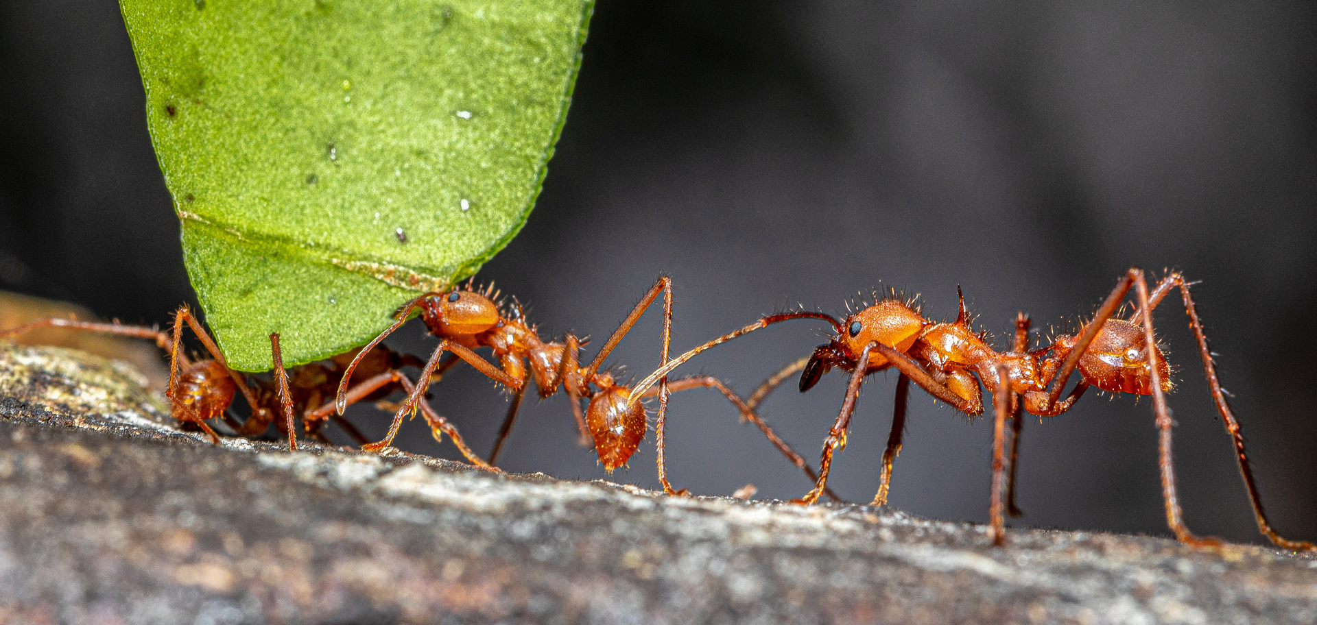
Worker leafcutter ants are separated into four, distinct, physical castes to handle specific jobs. Their brains, however, exhibit neuroplasticity in assigned roles.
Recently, researchers at the University of Pennsylvania revealed key elements of the leafcutters’ genetic code, pinpointing two signaling molecules that can be dialed up or down to reprogram ant duties. Crustacean cardioactive peptide (CCAP), elevated in Media ants, promotes leaf-harvesting tasks and can induce similar behavior in other subcastes. Neuroparsin-A (NPA), abundant in Majors, however, suppresses brood care and is linked to defensive patrols. Lowering the levels of NPA prompts caregiving behavior.
Reporting their findings in the journal Cell in July 2025, the researchers also uncovered a striking parallel: gene-expression patterns that govern division of labor in leafcutter ants mirror those in eusocial naked mole-rats—mammals that similarly abide by cooperative brood care—hinting at a convergent molecular mechanism dating back more than 600 million years. These results in ants reinforce how single neuropeptides can dramatically alter behavior, which may be applicable to human social behavior; although, humans are much more complex.
Building on previous research on carpenter ants, which have a similar but simpler social organization consisting of two roles (forager and soldier), the University of Pennsylvania team investigated several different neuropeptides in leafcutters to explore how these principles scaled up to the more nuanced, four-subcaste structure of Major, Media, Minor and Minima. They created 3D-printed, behavioral chambers that allowed them to monitor how ants interacted with the brood, fungus or leaves. These chambers enabled tracking and quantification of behavior through video analysis, demonstrating how altering CCAP and NPA levels in ants induced dramatic and reproducible shifts in their assigned tasks.
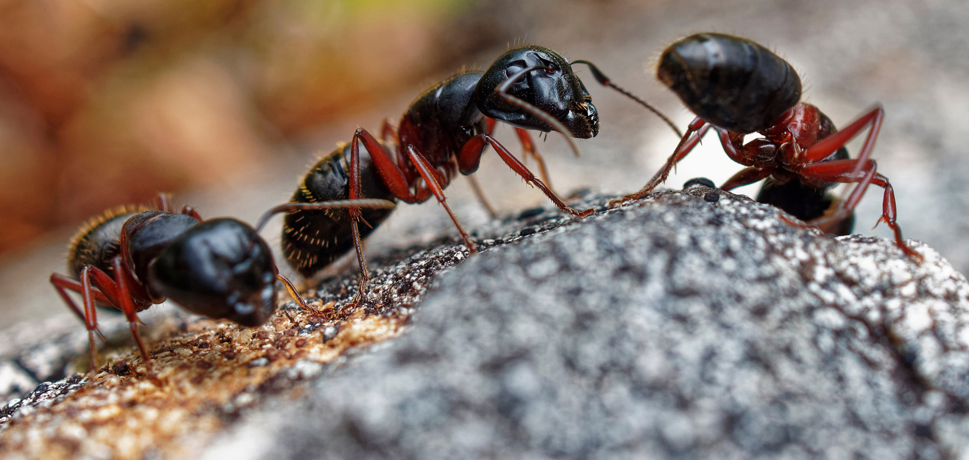
In contrast to leafcutter ants, carpenter ants have a simpler social organization consisting of two roles—forager and soldier—which is highly flexible. By manipulating core molecular pathways, researchers have scaled up their understanding of carpenter ants to reprogram behaviors within the leafcutter ant’s more intricate structure.
Generally speaking, say the scientists, specific neuropeptides are more abundant in certain castes. In Majors, the neuropeptide that dictates leafcutter behavior is low and the neuropeptide that stops nursing is high; so, if the former is increased in the ants’ brains, it leads to leafcutter behavior. If the latter is decreased, it leads to nursing and caretaking of the brood.
To further explore the evolutionary depth and implications of this behavioral script, the team investigated naked mole-rats, a distant evolutionary cousin whose subterranean colonies echo the caste-like harmony of leafcutter nests. Despite naked mole-rats lacking the exact neuropeptides (like NPA) found in ants, the researchers hypothesized that these peptides might still activate ancient, conserved pathways common to both species due to promiscuity of certain, more conserved receptors and convergent evolution. Remarkably, they found substantial overlap; enough that the ant neuropeptide could even activate endogenous receptors in the naked mole-rat brains.
Looking ahead, the scientists are keen to explore the persistence of biological plasticity as it relates to behaviors. They are also interested in extending their work to late-life rejuvenation and lifespan plasticity because reproductive ant queens live much longer than workers that don’t reproduce. They state that epigenetics, the study of how gene activity can be turned on or off without changing the underlying DNA, offers powerful ways to understand not only behavioral plasticity but lifespan pliability, too.
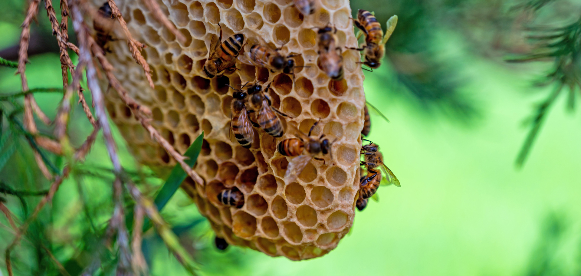
A new study offers insight into how a bee colony organizes its workforce without a leader assigning tasks. A hidden neural switch may determine whether a bee guards the hive, nurses the queen or searches for food.
Discovering the brain switch that assigns bees their jobs
Any successful community depends on its members completing the right jobs at the right time. Humans can discuss responsibilities and deliberately assign them, but a bee colony has no central planner. Even so, bees (Apis mellifera) divide their work with remarkable efficiency.
Worker bees’ responsibilities typically change as they grow older. Young workers care for the queen and developing larvae. Later, they help build and maintain the hive and defend it against threats. Only during the final stage of their lives do they leave the colony to search for food.
These behavioral changes are directed by interactions among roughly 1 million neurons in the bee brain. Until now, however, scientists did not understand how the nervous system produces this age-dependent progression from one task to another. Important clues emerged, however, from earlier research by a team at Germany’s Heinrich Heine University Dusseldorf (HHU). The scientists were studying a gene known as doublesex when they observed an unusual behavioral change. When the gene was deactivated in older worker bees, the insects began caring for the queen again. This responsibility is normally performed only by younger bees, suggesting that doublesex plays an important role in controlling age-related work behavior. Because the doublesex gene operates only within certain neural circuits, the discovery gave the HHU team and colleagues a way to investigate how specific parts of the bee brain influence social behavior.
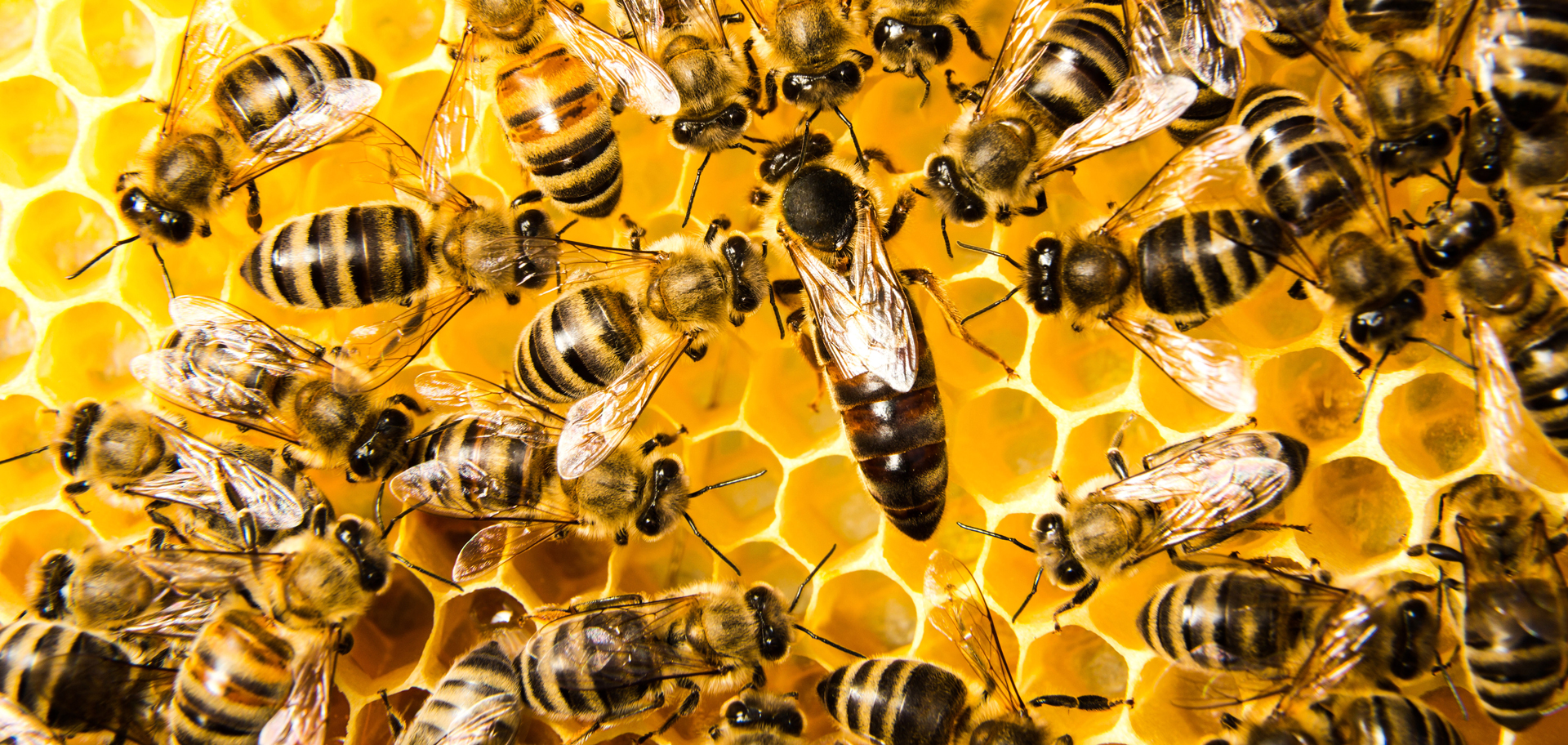
A queen bee is cared for and fed by young worker bees. As these bees get older, they take on other tasks in the hive. But biologists have found that they could change these behaviors by manipulating a particular gene and selectively reducing activity in certain neural circuits.
The researchers selectively silenced neurons associated with doublesex. To accomplish this, they used the gene to produce a protein that suppresses neural activity. They then activated that protein by feeding the bees a particular substance, allowing them to reduce activity only in the targeted circuits. Once those circuits were inhibited, older workers returned to caring for the queen instead of performing the duties expected at their age.
The results indicate that task organization within a bee colony has a neural foundation. When activity in particular areas of the brain is reduced, other neural circuits may become active and trigger a different set of behaviors. The findings provide early evidence that communication between neural circuits helps determine whether a worker bee nurses the queen, builds or guards the hive, or searches for food.
This ability to control the social behavior of bees offers new opportunities to explore the fundamentals of innate behavioral diversity and social cooperation, conclude the researchers in their report published in the journal Proceedings of the National Academy of Sciences in July 2026. The solution to the secret of how bees and other animals cooperate so well without a blueprint is likely hidden in the brain’s neural circuits.

It’s estimated that adults consume about 250 grams of microplastics each year. Some can accumulate in organs, including the brain. They may be quietly triggering brain inflammation and damage linked to Alzheimer’s disease and Parkinson’s disease.
Damaging your brain with microplastics
Dementia already affects more than 57 million people worldwide, and the number of individuals diagnosed with Alzheimer’s and Parkinson’s disease is expected to climb significantly in the coming years. Scientists say the possibility that microplastics could worsen or speed up these disorders raises serious public health concerns.
It’s estimated that adults consume about 250 grams of microplastics each year, roughly the amount needed to cover a dinner plate. Those microplastics come from a wide range of sources, including drinks in plastic bottles, food grown in contaminated soil, plastic cutting boards, processed foods, salt, contaminated seafood and tea bags, as well as plastic fibers from carpets, dust and synthetic clothing. Common plastics include polyethylene, polypropylene, polystyrene and polyethylene terephthalate or PET. The majority of these microplastics are cleared from our bodies; however, studies show they do accumulate in our organs, including our brains.
The findings come from a systematic review published in the journal Molecular and Cellular Biochemistry in November 2025. The research was carried out through an international collaboration led by scientists from Australia’s University of Technology Sydney and Alabama’s Auburn University. The scientists identified five key biological pathways that allow microplastics to harm the brain: activating immune cells, damaging neurons, disrupting the blood-brain barrier, increasing oxidative stress and interfering with mitochondria.
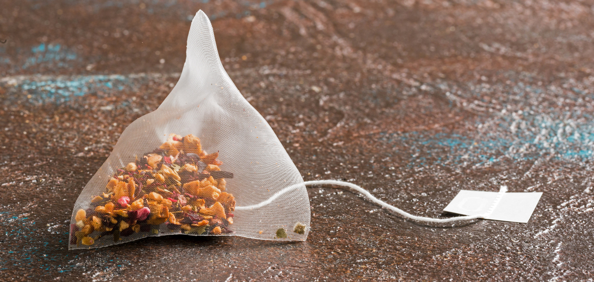
Luxury silk-style or pyramid-shaped tea bags are often made of nylon, PET or polypropylene, which can release billions of microplastic particles directly into your cup when exposed to boiling water. Even traditional paper tea bags advertised as natural wood fiber often use a polypropylene plastic lining or heat-sealing glue along the edges to hold their shape.
Microplastics weaken the blood-brain barrier, making it leaky. Once that happens, immune cells and inflammatory molecules are activated, which then causes even more damage to the barrier’s cells. The body treats microplastics as foreign intruders, which prompts the brain’s immune cells to attack them. When the brain is stressed by factors like environmental pollutants or toxins, this also creates oxidative stress in two primary ways: increased levels of “reactive oxygen species,” unstable molecules that can damage cells; and weakened antioxidant defenses that normally keep these molecules under control.
Microplastics also interfere with the way mitochondria produce energy, reducing the supply of ATP, or adenosine triphosphate, which is the fuel cells need to function. This energy shortfall degrades neuron activity and can ultimately damage brain cells. All these pathways interact with each other to increase damage in the brain.
The review also describes how microplastics might contribute to specific neurodegenerative diseases. In Alzheimer’s disease, they may promote the buildup of beta-amyloid and tau proteins. In Parkinson’s disease, they could encourage the aggregation of alpha-synuclein and harm dopaminergic neurons.

Classic fleece is made from polyester, a synthetic plastic material. Fleece clothing and blankets shed hundreds of thousands of microscopic plastic fibers into the air and water during normal wear and washing. If possible, choose natural fibers instead of synthetic ones.
Although current evidence suggests microplastics could worsen conditions such as Alzheimer’s and Parkinson’s, the authors emphasize that additional studies are needed to confirm a direct causal link. Even so, they recommend practical steps to reduce everyday exposure: use less plastic, steer clear of plastic containers and plastic cutting boards, don’t use the dryer, choose natural fibers instead of synthetic ones, and eat less processed and packaged foods.
Aggravating brain conditions through climate change
On top of the damage climate change has already had on the world, there is clear evidence that some brain conditions have also been affected, especially strokes and infections of the nervous system. Following a review of 332 papers published across the world between 1968 and 2023, scientists from the University College London said they expect the scale of the potential effects of climate change on neurological diseases to be substantial. They considered 19 different nervous system conditions, chosen on the basis of the 2016 Global Burden of Disease study, including Alzheimer’s, epilepsy, meningitis, migraine, multiple sclerosis and stroke. The impact of climate change on several serious but common psychiatric disorders, including anxiety, depression and schizophrenia, were also analyzed.
The climatic variations that were shown to have an effect on brain diseases included extremes of temperature (both high and low) and greater temperature swings throughout the course of day, especially when these measures were seasonally unusual. Nighttime temperatures may be particularly important, as higher temperatures through the night can disrupt sleep. Poor sleep is known to aggravate a number of brain conditions. The researchers found that there was an increase in health-care facility admissions, disabilities and mortalities as a result of strokes in higher ambient temperatures or during heat waves.

There is an urgent need to understand the impact of climate change on people with neurological conditions in order to preserve their health. For example, higher nighttime temperatures can disrupt sleep, aggravating a number of brain conditions, including Alzheimer’s, dementia, migraines and Parkinson’s. ©davide bonaldo/Shutterstock.com
Meanwhile, state the researchers in their paper published in the journal The Lancet Neurology in June 2024, people with dementia are additionally susceptible to harm from extremes of temperature (such as heat-related illnesses or hypothermia) and weather events (like flooding or wildfires) because cognitive impairment can limit their abilities to adapt their behaviors to environmental changes. A reduced awareness of risks might be combined with a diminished capacity to seek help or to mitigate potential harm, such as by drinking more in hot weather or by adjusting clothing. Climate anxiety is an added, potentially weighty influence: many brain conditions are associated with higher risk of psychiatric disorders, including anxiety.
Triggering brain healing in virtual nature
A growing body of evidence is showing that exposure to nature can help reduce short-term, everyday pain, but there has been less research into how this might work for people living with chronic or longer-term pain. Chronic pain typically lasts more than three months and is particularly difficult to treat. Now, a group of researchers led by those at England’s University of Exeter has tested the impact of immersive, 360-degree nature films delivered using VR compared with 2D video images in reducing the experience of pain. The findings of their study, published in the journal Pain in September 2025, show that virtual reality is almost twice as effective, performing similar to painkillers.
The study involved 29 healthy participants who were shown two types of nature scenes after having pain delivered on the forearm using electric shocks. On the first visit, in the absence of any nature scenes, the scientists measured the changes in pain that occurred over a 50-minute period following the electric shocks by delivering sharp, pricking stimuli. The results showed that the participants developed a type of sensitivity that closely resembles that seen in people living with nerve pain.

When people were given a 45-minute, 360-degree virtual reality experience of the waterfalls of Oregon, their feelings of pain sensitivity were significantly reduced.
On the second visit, they immersed the same participants in a 45-minute, virtual reality, 360-degree experience of the waterfalls of Oregon to test for any changes in the development of pain sensitivity. In a third visit, they saw the same scene, but on a 2D screen. They then completed questionnaires on their experiences of pain after watching the scenes in each case, on how present they felt in each experience and to what extent they felt the nature scenes to be restorative.
On a separate visit, participants underwent MRI brain scans at the University of Exeter. Researchers administered a cold gel to illicit a type of ongoing pain and then scanned participants to study how their brains responded. The researchers found that the immersive VR experience significantly reduced the development and spread of feelings of pain sensitivity to pricking stimuli, and these pain-reducing effects were still there even at the end of the 45-minute experience.
The more present a person felt during the VR experience, the stronger this pain-relieving effect, suggesting that nature scenes delivered using VR can help to change how pain signals are transmitted in the brain and spinal cord during long-term pain conditions. The University of Exeter researchers hope that this study leads to more research into how exposure to nature affects our pain responses and that nature scenes will be further incorporated into home-care settings and hospitals.

Not everyone is able to get outside for walks in nature, particularly those living with long-term health conditions like chronic pain. However, virtual reality nature experiences can trigger the brain’s natural painkillers, reducing pain sensitivity almost as effectively as medication. Shown here is a 3D, spherical panorama with a 360-degree viewing angle.
Easing brain pain
The brain craving shown by the zombies in the 1985 movie Return of the Living Dead was an effort to ease their pain. According to zombie lore, the state of being dead and decomposing is intensely hurtful for zombies. Eating fresh human brains temporarily dulls that persistent pain.
I have a thought. Instead of scaring us by lusting for fresh brains, the zombies might want to try immersing themselves in nature, whether in the real world or via virtual reality.
Here’s to finding your true places and natural habitats,
Candy