


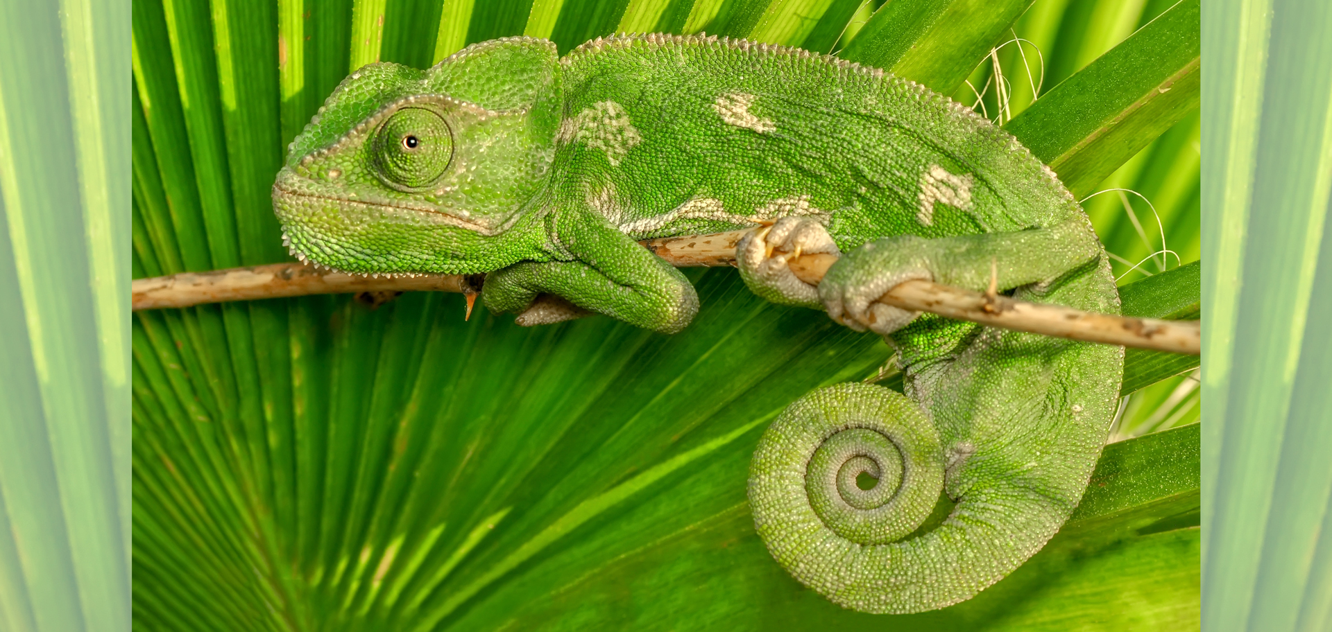
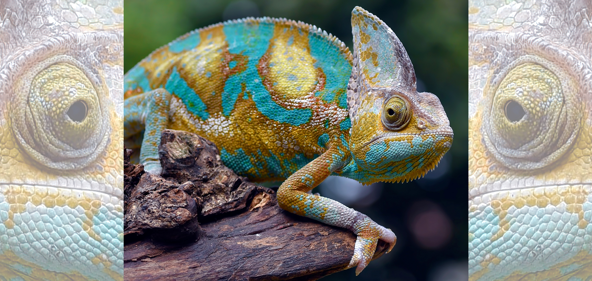
Chameleons have the extraordinary ability to move their eyes independently, stemming from an anatomical marvel: long, tightly coiled optic nerves hidden behind their bulging eyes. The coils give the eyes extra slack, enabling nearly 360-degree scanning without neck mobility. This configuration is not documented in any other lizard species.
Eyes are our primary sensory organs, allowing us to perceive about 80% of incoming information, to navigate safely and to perform daily tasks like reading and work, while also acting as vital indicators for our overall physical health. They’re also central to our nonverbal communication. Changes in expression, focus and pupil size reveal our emotional states, such as crinkling our eyes in a true smile or enlarging the size of our pupils in response to having strong feelings, such as attraction, excitement or fear. People naturally look at our eyes during social interactions, making them a primary channel for interpreting emotions and spotting honesty.
However, we’re not the only creatures with fascinating eyes. Chameleons have the extraordinary ability to move their eyes independently. Golden apple snails (Pomacea canaliculata) can fully regrow their eyes; and with genes and eye structures that are strikingly similar to ours, that raises hope for future human-vision restoration.
And then there’s the soulful eyes of our pet dogs. They reflect a deep, unspoken bond, conveying loyalty, unconditional love and understanding through a gaze that triggers the release of oxytocin in both them and us. This profound connection feels like a glimpse into dogs’ pure, nonjudgmental world, offering comfort and a sense of being truly seen. But it may humble and surprise you to learn that “puppy dog eyes” didn’t evolve exclusively in dogs because of domestication. Those famous eyes are, in fact, shared with some of their wild cousins.

A chameleon can extend its tongue up to two body lengths, launching from zero to 60 miles per hour in about a hundredth of a second, with accelerations exceeding 250 Gs, one of the fastest movements for snatching prey in the animal kingdom.
Solving a 2,000-year-old mystery about chameleon eyes
Chameleons inhabit parts of Africa, Asia and Europe, and their remarkable adaptations go far beyond color change. They move through trees using a prehensile tail for balance and mitten-like feet for a careful, measured stride. Their slow pace is compensated for by a high-speed weapon: a tongue that can accelerate from zero to 60 miles per hour in about one-hundredth of a second. This sticky, elongated tongue can strike prey located at more than double the chameleon’s own body length.
But for thousands of years, it’s the eyes of chameleons that have most intrigued observers and scientists. While scanning their environment to find prey, the animals move their eyes independently, in all directions, almost like security cameras. But the moment that they find their prey, their eyes coordinate and work together in one direction to calculate where to shoot their tongues.
With such impressive qualities, it is unsurprising that chameleons have appeared in human culture for millennia. Their recognizable silhouettes, complete with coiled tails, are even present in ancient Egyptian rock carvings.
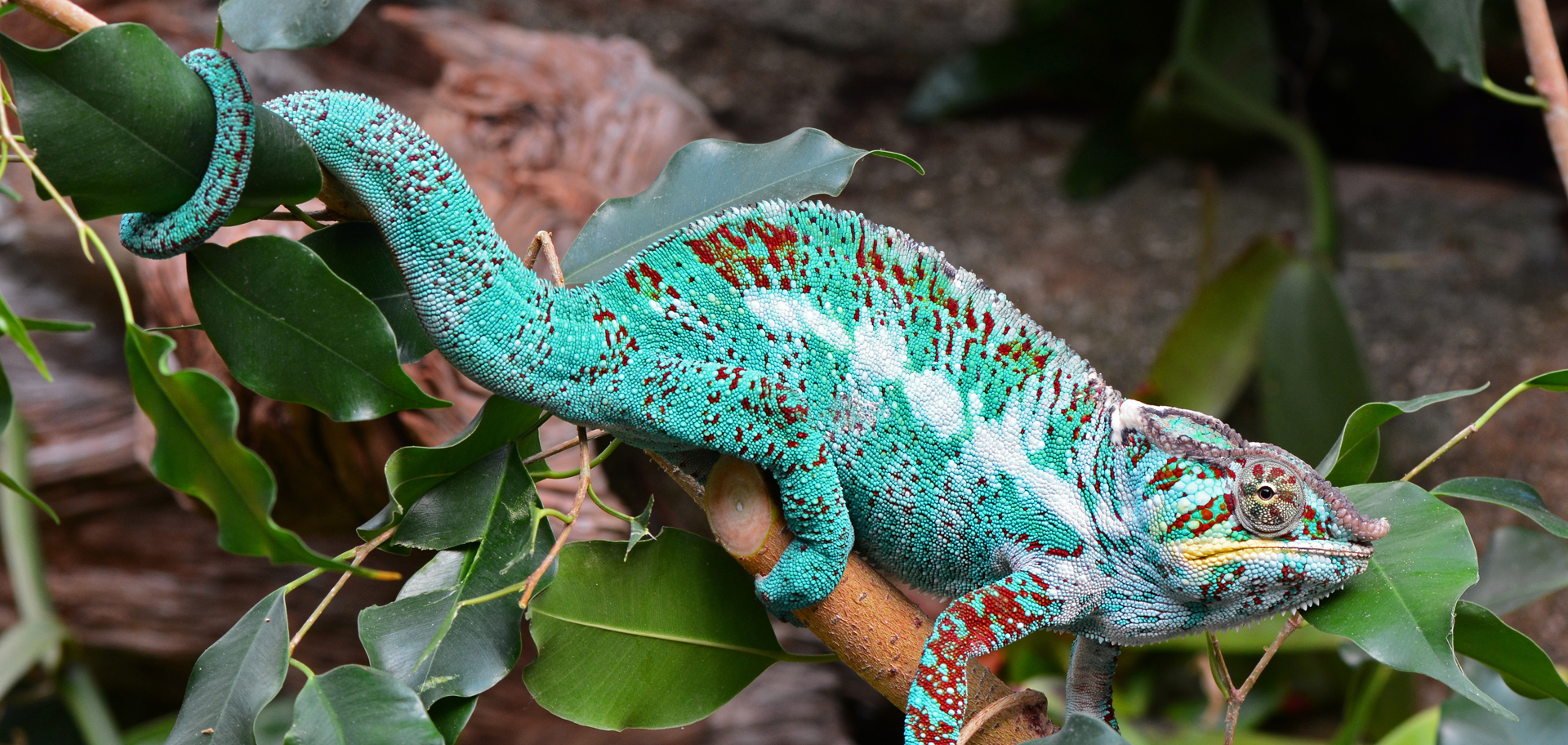
Chameleons have prehensile tails, meaning that they’re able to grasp and hold onto objects. In the wild, these lizards live most of their lives in the trees and use their tails to help them climb and maintain their balance while they’re walking on thin branches.
More than 2,000 years ago, Greek philosopher Aristotle incorrectly suggested that chameleons entirely lacked optic nerves. He believed their eyes were connected directly to the brain, which, in his view, explained their independent movement. In the mid-1600s, Roman physician Domenico Panaroli refuted this idea, asserting that chameleons do possess optic nerves, but that they do not cross as they do in many other animals. In most vertebrates, such crossing transfers information from the right eye to the left side of the brain and vice versa. Panaroli reasoned that the absence of this crossing granted chameleons greater freedom of eye movement.
English mathematician and physicist Isaac Newton later supported Panaroli’s conclusions. He referenced chameleons in his 1704 book Optiks, a collection of three decades of his ideas on color and light. However, French anatomist Claude Perrault had already drawn a much more accurate representation in 1669, showing two optic nerves that crossed and then continued straight. His illustration received little attention from Newton’s contemporaries, even though it was one of the clearest early depictions.
Over time, published diagrams came close to showing the true shape of the optic nerves but never captured it fully. Johann Fischer’s 1852 treatise on lizard neuroanatomy included part of a curled structure but omitted the rest; and in 2015, a master’s degree student at Israel’s University of Haifa noted a C-shaped section of the optic nerves. But no complete description existed in the scientific literature.
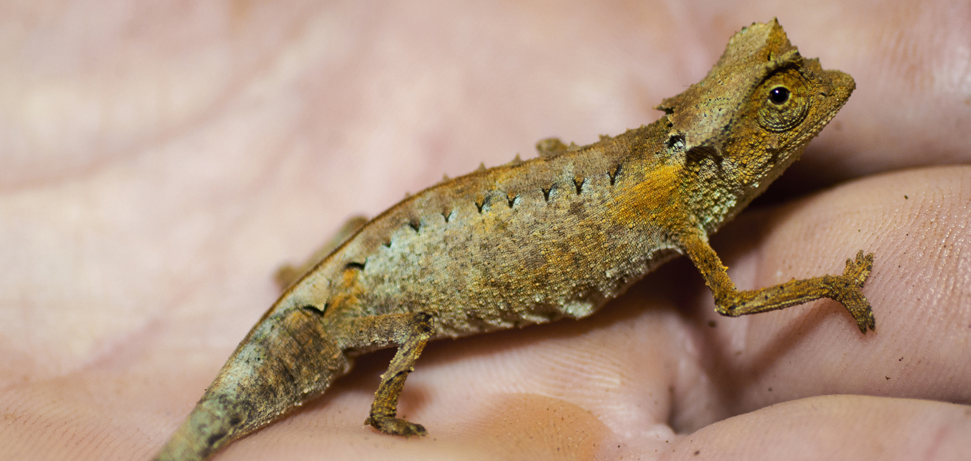
Tiny minute leaf chameleons aren’t the usual, brightly colored chameleons. Dark brown on the limbs and brown with black splotching on the sides, they blend in along the low branches and leaf litter of their forest habitats.
So, although chameleons’ shifting gaze is easy to observe, the internal structures enabling it have remained unclear. Then, in 2017, Edward Stanley, director of the Florida Museum of Natural History’s digital imaging laboratory, noticed an unexpected pattern: a CT scan of the minute leaf chameleon (Brookesia minima) revealed tightly coiled optic nerves, a shape unlike anything he had ever encountered before.
That spurred a research team, led by the museum and involving 18 other U.S. institutions, to comb through vast archives of 3D digital models of vertebrate anatomy. They enlisted language experts to interpret old anatomical works written in French, Italian and Latin, and sometimes in a perplexing blend of several languages. How such a distinctive feature remained hidden for so long became clear as the scientists examined historical research methods. Earlier studies relied heavily on physical dissections. These procedures frequently damaged or shifted the fragile optic nerves, making accurate observations nearly impossible.
Today, CT scanning is widespread in medical and scientific settings. High-resolution, X-ray CT makes it possible to view structures concealed inside preserved specimens, including the interior of a chameleon’s skull. Spotting a coiled optic nerve in one chameleon provided an important clue, but researchers needed broader evidence. Fortunately, they had access to extensive digital resources through oVert (short for openVertebrate).
Veiled chameleons are native to the Arabian Peninsula in Saudi Arabia and Yemen. They occupy a variety of habitats, including dry and high plateaus, forests and river valleys. They are among the most popular chameleon species kept as pets.
Using oVert datasets, CT scans from more than 30 lizards and snakes were examined, including three chameleon species representing major lineages. The researchers built 3D brain models for 18 of these reptiles and measured the optic nerves in each. All three chameleon species displayed optic nerves that were significantly longer and more tightly coiled than those of the other lizards. This confirmed that the initial finding in 2017 was representative of the group.
The researchers then investigated how the coils develop in young chameleons. Examining embryos of the veiled chameleon (Chamaeleo calyptratus) at three stages, they noted that the optic nerves start straight and lengthen over time, eventually forming loops before the animal hatches. Hatchlings already possess fully mobile eyes.
Determining when this feature evolved is more difficult. The oldest known chameleon fossils date to the early Miocene, about 16 to 23 million years ago, long after many of their arboreal adaptations had appeared. While these fossils don’t reveal much about the sequence in which traits emerged, the newly documented nerve coils provide a clue about why this adaptation may have arisen.
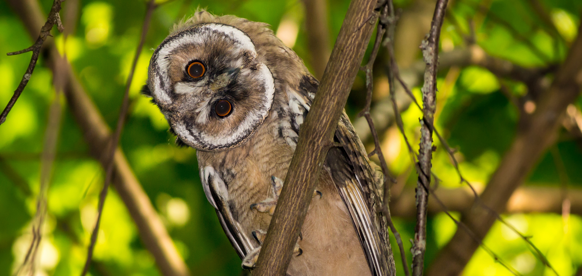
Owls can turn their heads up to 270 degrees due to extra neck bones, a single pivot joint allowing free rotation, large air sacs that cushion arteries and special blood vessel adaptations that prevent damage and maintain blood flow to their fixed, tubelike eyes, which can’t swivel.
Many vertebrates with large eyes expand their field of view in one of two ways: by turning their head or by moving their eyes extensively. Owls and lemurs rotate their necks to look around. Humans and some other mammals rely on stretchy optic nerves that allow substantial eye movement. Rodents achieve a similar effect with wavy nerve fibers that add flexibility.
Chameleons, however, do not have flexible necks. The coiled optic nerve could have developed as a work-around, giving the eyes extra slack and reducing strain as they pivot. A comparable adaptation has been noted in only a few invertebrates, such as the stalk-eyed fly.
In their study’s conclusion, published in the journal Scientific Reports in November 2025, the scientists compare the optic nerves of chameleons with old phones. The first phones had just simple, straight cords attached to the headsets, but then someone had the idea to coil the cords and give them more slack so people could walk farther with them. That’s what the chameleons are doing: they’re maximizing the range of motion of their eyes by creating coiled structures. Next, the study’s authors hope to discover whether other tree-dwelling lizards evolved similar solutions.
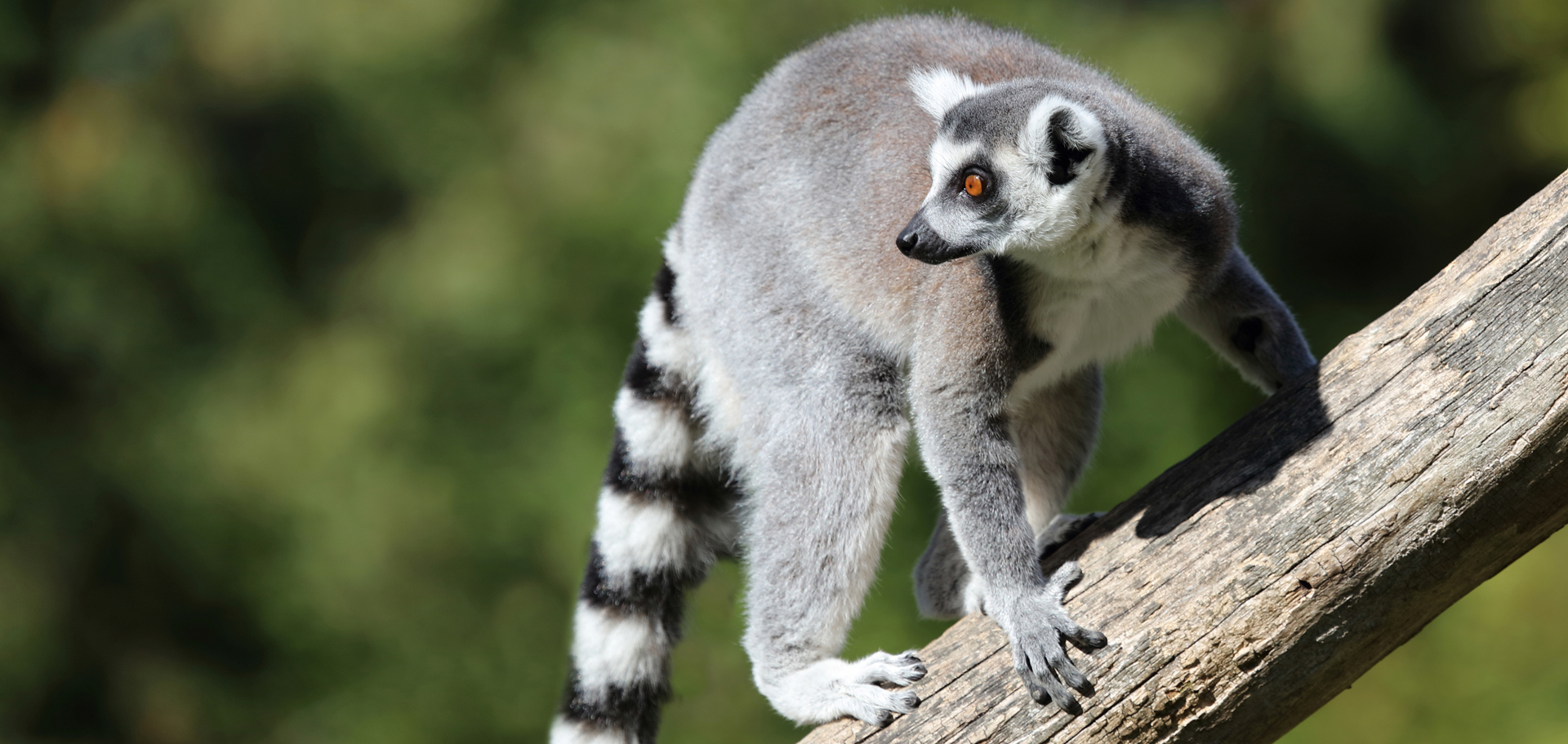
Similar to other primates, lemurs turn their heads using typical neck-joint movements. Unlike animals such as owls whose eyes are fixed and thus must rotate their heads to see, lemurs have forward-facing eyes that can move to some extent, allowing for stereoscopic vision.
Regrowing snail eyes
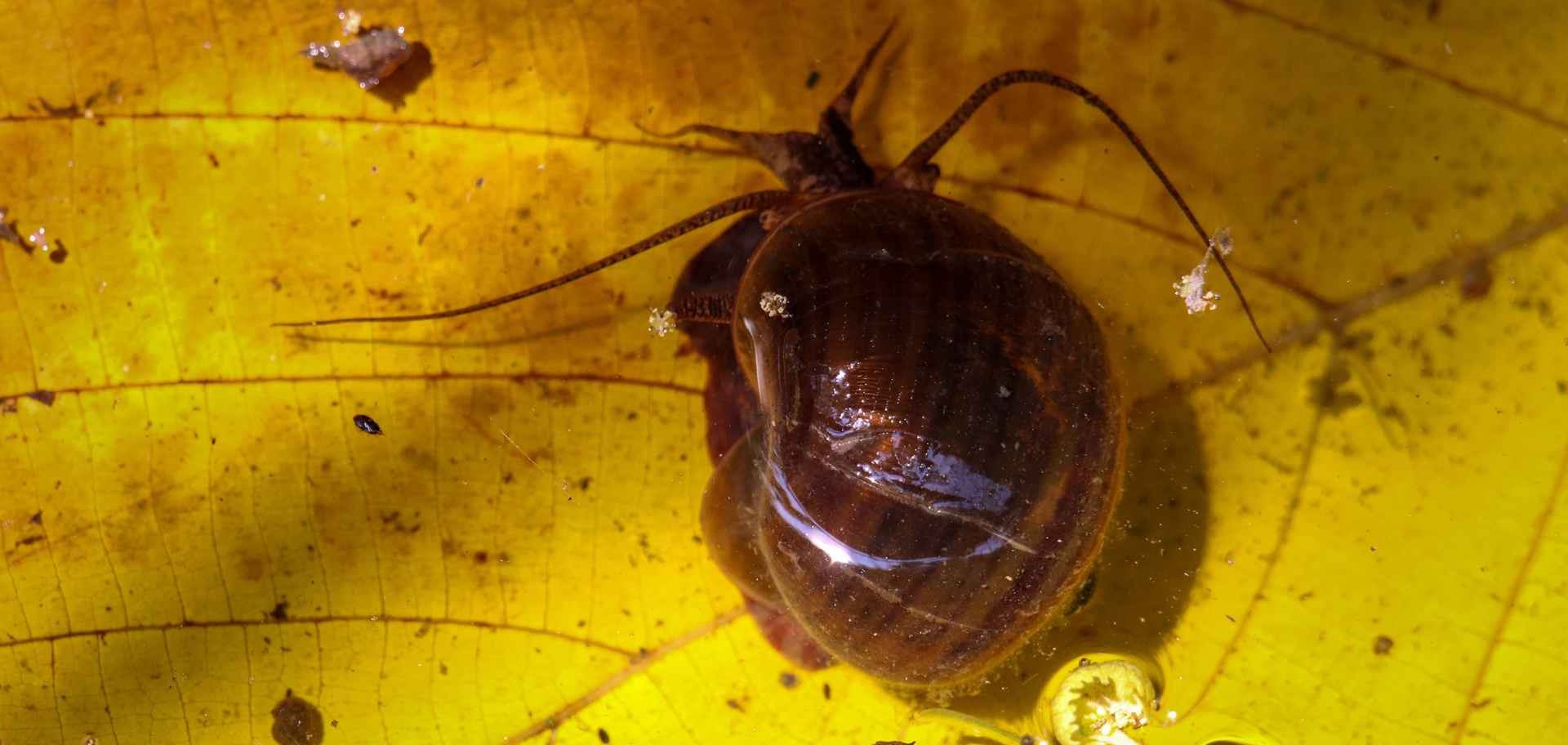
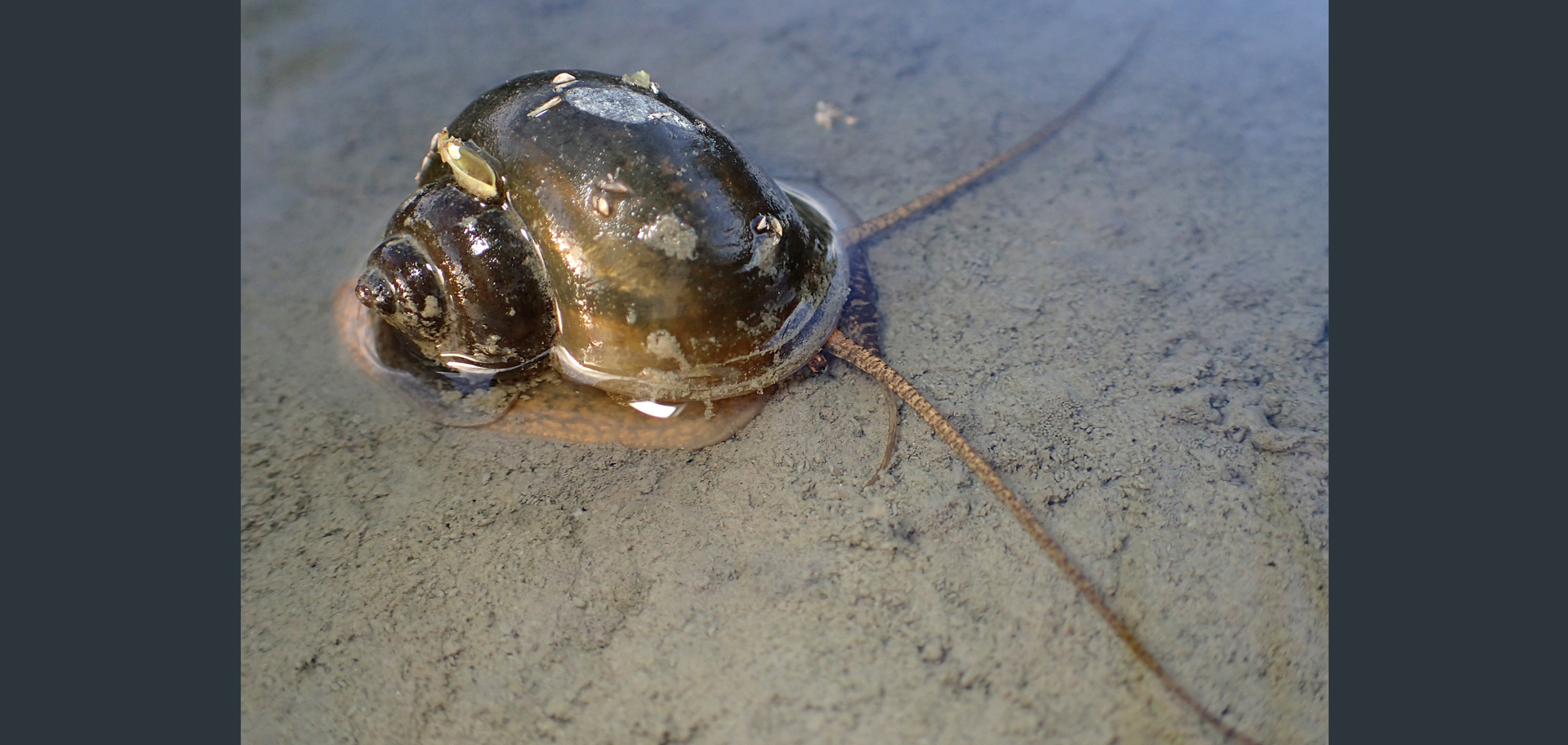
Human eyes are complex and intricate—and they’re irreparable. Structurally, though, they are comparable to those of the freshwater golden apple snail, an animal which can completely regenerate its eyes.
In a study published in the journal Nature Communications in August 2025, biologists at the University of California, Davis, show that golden apple snail and human eyes share many anatomical and genetic features. They hope to shed light on how these snails regrow their eyes, with the goal of eventually helping to restore vision in people with eye injuries. The team also developed methods for editing the golden apple snail’s genome, which will allow them to explore the genetic and molecular mechanisms behind eye regeneration.
Snails have been known for their regenerative abilities for centuries. In 1766, a researcher noted that decapitated garden snails can regrow their entire heads. Nevertheless, the University of California, Davis, scientists are the first to leverage this feature in regenerative research.
The golden apple snail is characterized by fast growth, a high reproduction rate, strong stress tolerance and adaptation to a broad range of environments. Native to South America, golden apple snails are now invasive in many places throughout the rest of the world. ©Chung Yi Fang/Shutterstock.com
Although native to South America, golden apple snails are now invasive in many places throughout the rest of the world. But the same traits that make the snails so invasive also make them a good animal to work with in labs. They’re resilient, their generation time is very short, and they have a lot of babies. In addition, they have “camera-type” eyes—the same kind as humans do.
There are many types of eyes in the animal kingdom, but camera-type eyes are known for producing particularly high-resolution images. They consist of a protective cornea, a lens for focusing light and a retina that contains millions of light-detecting photoreceptor cells. They are found in all vertebrates, some spiders, octopuses and squid, and some snails.
Using a combination of genomic analysis, microscopy and other scientific techniques, the biologists demonstrated that the golden apple snail’s eyes are anatomically and genetically similar to human eyes, and that many genes that participate in human eye development are also present in the snail. And after regeneration, the morphology and gene expression of a new snail eye is almost identical to the original one.
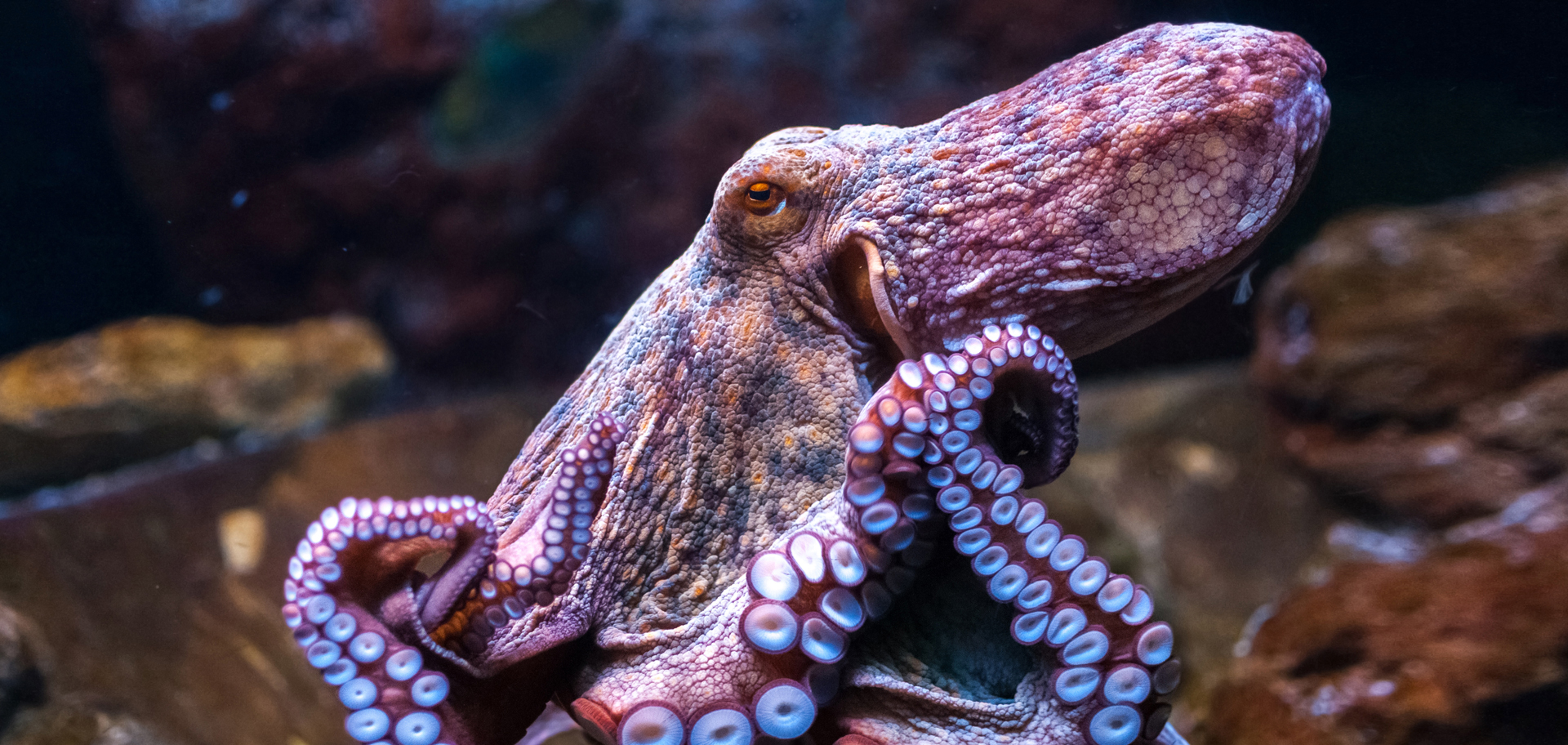
Octopuses have complex, camera-type eyes, similar in structure and function to human eyes; though they focus their vision by physically moving their rigid lenses forward or backward rather than changing the shape of their lenses like humans do. In addition, they have a unique visual system adapted for their marine environment.
It takes about a month for a snail to grow a new eye and it’s done in several phases. First, the wound must heal to prevent infection and fluid loss, which usually takes about 24 hours. Then, unspecialized cells migrate and proliferate in the area. Over the course of about a week and a half, these cells specialize and begin to form eye structures including the lens and retina. By the 15th day after eye loss, all of the eye’s structures are present, including the optic nerve, but these structures continue to mature and grow for several more weeks.
Although the biologists don’t have conclusive evidence yet that the snails with regrown eyes can see images, anatomically they have all the components that are needed to form an image. They’re now working on a behavioral assay to show that the snails can process stimuli using their new eyes in the same way as they did with their original eyes.
The team also investigated which genes were active during the regeneration process. They showed that immediately after the eye loss, the snails had about 9,000 genes that were expressed at different rates compared to normal adult snail eyes. After 28 days, 1,175 genes were still expressed differently in the regenerated eye, which suggests that although the eyes look fully developed after a month, complete maturation might take longer.
Unlike us, golden apple snails can regenerate a damaged or missing eye. Biologists are studying how the snails accomplish this feat, knowledge that could help us understand eye damage in humans—and even lead to new ways to heal or regenerate human eyes.
The University of California, Davis, scientists are also exploring other eye-related genes, including genes that encode specific parts of the eye, like the lens or retina. If they find a set of genes that are important for eye regeneration and these genes are also present in vertebrates, in theory they could activate them to enable eye regeneration in humans.
Sharing puppy dog eyes
While chameleons can move their eyes in almost all directions and snails can regrow theirs, there probably isn’t a set of eyes that touch our hearts more than puppy dog eyes.
However, “puppy dog eyes” may be a misnomer. New research from Baylor University in Texas reveals that coyotes, like domestic dogs, can produce the famous expression formerly ascribed to our “best friends” only. Publishing their findings in the journal Royal Society Open Science in October 2024, the university authors challenge the hypothesis that this facial feature evolved exclusively in dogs as a result of domestication.

Oh, those puppy dog eyes! When dogs raise their inner eyebrows, it makes their eyes appear larger and more infant-like, eliciting a nurturing response from humans. This gesture has evolved over time as a nonverbal method to communicate and bond with those around them.
The muscle responsible for raising the inner eyebrow to create puppy dog eyes is the levator anguli oculi medialis (LAOM). The Baylor University researchers discovered that coyotes also possess a well-developed LAOM, like that of dogs. This finding contradicts the idea that the muscle evolved specifically for communication between humans and dogs during domestication. Rather, it’s an ancestral trait shared by multiple species in the Canis genus, raising intriguing questions about the role of facial expressions in communication and survival among wild canids.
To reach their conclusions, the scientists compared the facial muscles of coyotes, dogs and gray wolves. While both dogs and coyotes possess a well-developed LAOM, the muscle is either modified or absent in gray wolves. This means that human-driven selection wasn’t solely responsible for the development of the inner brow-raiser in dogs. Instead, the LAOM was likely present in a common ancestor of dogs, coyotes and gray wolves but was later lost or reduced in wolves.
The research also documented significant, intraspecific variation in the facial muscles of coyotes, particularly those related to brow and lip movements. Genetic analysis was used to rule out significant dog ancestry in the coyote participants, reinforcing that these traits are not a result of crossbreeding. This work shows that coyotes and dogs share not just behavioral similarities, but also an engaging evolutionary history that includes the ability to make expressions that we once thought were unique to domesticated animals.
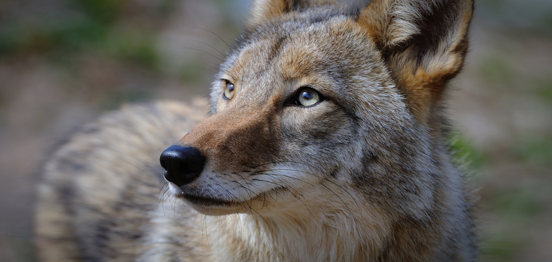
Dogs have a specific muscle—the LAOM—around their eyes that allows them to make animated expressions. Scientists just learned that coyotes also possess a well-developed LAOM that’s similar to that of dogs.
This discovery has broader implications for understanding the evolution of facial expressions in mammals, state the researchers. The LAOM may have originally evolved for functions related to eye movements and vision, rather than communication with humans, as once thought. Future studies on other canid species, including African wild dogs and red wolves, may further illuminate the role of facial expressions in survival and species communication.
Feeling forest eyes
Writing about eyes puts me in mind of one of the most beautiful statements written about them that I have come across. Anthropologist, author and storyteller Richard Nelson wrote, “I am never alone in this wild forest, this forest of elders, this forest of eyes.”
The sentence appeared in his 1991 book The Island Within and conveys his deep relationship with a forest on a remote, unnamed island in the Pacific Northwest. In association with the Koyukon teachings he received, Richard Nelson believed that forests had a presence, wisdom and spiritual power that he could physically feel through what he could only describe as “eyes.”
At least once in your life, you’ve almost certainly felt a spiritual “forest of eyes”; especially if you’ve ever looked into the eyes of a wild “other.”
You’ve felt those kinds of eyes, too; every time you’ve looked into those of your loved ones—or the eyes of the wild “others” you’ve been lucky enough to hold for a brief moment.
Here’s to finding your true places and natural habitats,
Candy


